
Page 90 - Clinical Anatomy
P. 90
ECA2 7/18/06 6:42 PM Page 75
The gastrointestinal tract 75
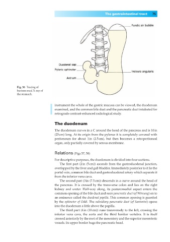
Fig. 56◊Tracing of
barium meal X-ray of
the stomach.
instrument the whole of the gastric mucosa can be viewed, the duodenum
examined, and the common bile duct and the pancreatic duct intubated for
retrograde contrast-enhanced radiological study.
The duodenum
The duodenum curves in a C around the head of the pancreas and is 10in
(25cm) long. At its origin from the pylorus it is completely covered with
peritoneum for about 1in (2.5cm), but then becomes a retroperitoneal
organ, only partially covered by serous membrane.
Relations (Figs 57, 58)
For descriptive purposes, the duodenum is divided into four sections.
The first part (2in (5cm)) ascends from the gastroduodenal junction,
overlapped by the liver and gall-bladder. Immediately posterior to it lie the
portal vein, common bile duct and gastroduodenal artery which separate it
from the inferior vena cava.
The second part (3in (7.5cm)) descends in a curve around the head of
the pancreas. It is crossed by the transverse colon and lies on the right
kidney and ureter. Half-way along, its posteromedial aspect enters the
common opening of the bile duct and main pancreatic duct (of Wirsung) on to
an eminence called the duodenal papilla. This common opening is guarded
by the sphincter of Oddi. The subsidiary pancreatic duct (of Santorini) opens
into the duodenum a little above the papilla.
The third part (4in (10cm)) runs transversely to the left, crossing the
inferior vena cava, the aorta and the third lumbar vertebra. It is itself
crossed anteriorly by the root of the mesentery and the superior mesenteric
vessels. Its upper border hugs the pancreatic head.