
Page 95 - Clinical Anatomy
P. 95
ECA2 7/18/06 6:42 PM Page 80
80 The abdomen and pelvis
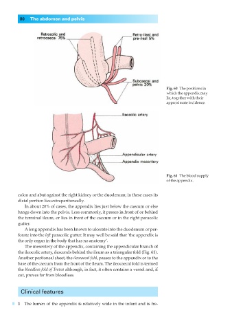
Fig. 60◊The positions in
which the appendix may
lie, together with their
approximate incidence.
Fig. 61◊The blood supply
of the appendix.
colon and abut against the right kidney or the duodenum; in these cases its
distal portion lies extraperitoneally.
In about 20% of cases, the appendix lies just below the caecum or else
hangs down into the pelvis. Less commonly, it passes in front of or behind
the terminal ileum, or lies in front of the caecum or in the right paracolic
gutter.
Along appendix has been known to ulcerate into the duodenum or per-
forate into the left paracolic gutter. It may well be said that ‘the appendix is
the only organ in the body that has no anatomy’.
The mesentery of the appendix, containing the appendicular branch of
the ileocolic artery, descends behind the ileum as a triangular fold (Fig. 61).
Another peritoneal sheet, the ileocaecal fold, passes to the appendix or to the
base of the caecum from the front of the ileum. The ileocaecal fold is termed
the bloodless fold of Treves although, in fact, it often contains a vessel and, if
cut, proves far from bloodless.
Clinical features
1◊◊The lumen of the appendix is relatively wide in the infant and is fre-