
Page 177 - Clinical Application of Mechanical Ventilation
P. 177
Special Airways for Ventilation 143
With each breath, the tube is advanced into the trachea until the bronchial seg-
ment plugs the bronchus. The endpoint signs are (1) resistance to advancement
(even with low compliance lungs), (2) unilateral ventilation by observation and
auscultation, and (3) reduction in compliance (e.g., cPIP in volume-controlled
ventilation). At this point, the tube is about 2.5 to 3 cm (bronchial cuff length plus
1 cm) from its final position.
Once the bronchial plugging point has been identified, the bronchial cuff is de-
flated and the tube is inserted another 2.5 to 3 cm (bronchial cuff length plus 1 cm).
The final bronchial cuff volume needed to seal should be small, about 1 to 2 mL.
The patient connection is now changed to the tracheal connection. The tracheal cuff
is inflated until a seal is made in the trachea. Proper positioning of the DLT can be
Proper positioning of verified by auscultation or fiberoptic bronchoscopy (Klein et al., 1998).
the DLT can be verified by
auscultation or fiberoptic If the left-sided tube goes right, turning the head 90º so that the chin is on the
bronchoscopy.
right shoulder and rotating the tube on its axis to restore the bronchial tube to
point laterally to the left can usually achieve insertion into the left main bronchus
(Russell, 2004).
Complications of DLT
Use of DLTs may lead to airway injuries (Campos et al., 2000), and the most
The most severe form of severe form of airway injuries is airway rupture. The incidence of airway rupture is
airway injuries caused by DLTs
is airway rupture. higher when large and medium-sized red rubber DLTs are used. In contrast, small
polyvinyl-chloride (PVC) DLTs have been associated with airway rupture (Akhtar,
1999). Some risk factors for airway rupture are listed in Table 5-9.
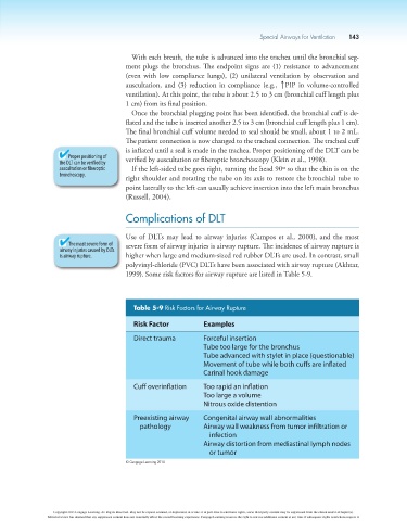
Table 5-9 Risk Factors for Airway Rupture
Risk Factor Examples
Direct trauma Forceful insertion
Tube too large for the bronchus
Tube advanced with stylet in place (questionable)
Movement of tube while both cuffs are inflated
Carinal hook damage
Cuff overinflation Too rapid an inflation
Too large a volume
Nitrous oxide distention
Preexisting airway Congenital airway wall abnormalities
pathology Airway wall weakness from tumor infiltration or
infection
Airway distortion from mediastinal lymph nodes
or tumor
© Cengage Learning 2014
Copyright 2013 Cengage Learning. All Rights Reserved. May not be copied, scanned, or duplicated, in whole or in part. Due to electronic rights, some third party content may be suppressed from the eBook and/or eChapter(s).
Editorial review has deemed that any suppressed content does not materially affect the overall learning experience. Cengage Learning reserves the right to remove additional content at any time if subsequent rights restrictions require it.