
Page 188 - Clinical Application of Mechanical Ventilation
P. 188
154 Chapter 6
COMMON ARTIFICIAL AIRWAYS IN
MECHANICAL VENTILATION
An ET tube may be inserted orally (oral intubation) or nasally (nasal intubation)
Oral intubation is easy to through the larynx into the trachea. Oral intubation is easy to perform and it is
perform and is often done in
emergency situations. often done in emergency situations. Nasal intubation is more time-consuming and
it is more suitable in elective intubations.
Some ET tubes (e.g., Spiral-Flex®) are reinforced with spiral stainless steel wire
within the tube wall to reduce risk of tube kinking. They are the ideal choice for
head and neck surgery when bending or compression of the tube is likely to occur, or
for patients in the ICU biting on the tube or experiencing seizures. These tubes are
part metal and must be replaced with a regular ET tube when the patient undergoes
any procedure involving magnetic resonance imaging.
Endotracheal Tube
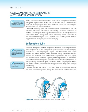
Intubation through the mouth is the preferred method of establishing an artificial
airway (Figure 6-3). An oral route provides quick access to the lungs in emergency
An oral route of intuba-
tion allows the passage of situations and it allows the passage of a larger ET tube than the nasal route. A larger
a larger ET tube than nasal tube has less airflow resistance, and it lowers the airway pressure requirements.
intubation.
However, oral intubation is less comfortable to the patient and may cause gagging
and excessive secretion production. Agitated patients may bite down on the tube and
cause airflow obstruction. In general, the oral route of intubation may be preferred for
cardiopulmonary resuscitation, apneic patient, nasal trauma, nasopharyngeal obstruc-
A larger ET tube lowers tion, midfacial trauma, basilar skull fracture, epiglottitis, and anticipation of fiberoptic
the peak, plateau, and mean
airway pressures. bronchoscopy.
Another common ET tube (e.g., Hi-Lo Evac) has an evacuation lumen/port
that allows continuous aspiration of subglottic secretions. Studies have shown that
© Cengage Learning 2014
Figure 6-3 This illustration shows how an inflated cuff seals the trachea.
Copyright 2013 Cengage Learning. All Rights Reserved. May not be copied, scanned, or duplicated, in whole or in part. Due to electronic rights, some third party content may be suppressed from the eBook and/or eChapter(s).
Editorial review has deemed that any suppressed content does not materially affect the overall learning experience. Cengage Learning reserves the right to remove additional content at any time if subsequent rights restrictions require it.