
Page 666 - Clinical Application of Mechanical Ventilation
P. 666
632 Chapter 19
The initial blood pressure was 139/100 mm Hg, temperature was 35.8°C with a
Cyanide is one of the by- pulse of 102/min, and respiratory frequency of 24/min while breathing on a non-
products of combustion.
rebreather mask at 15 L/min. He was immediately fluid-resuscitated with 1,000 mL
of D W, and treated empirically for cyanide toxicity, which interferes with oxidative
5
metabolism at the cellular level by impairing the utilization of oxygen in the tissues.
With adequate spontane- The clinical signs of cyanide poisoning at different toxic levels may include the
ous ventilation, the initial following:
supportive measure for smoke
inhalation is 100% O 2 .
Blood cyanide concentration 0.2 to 0.3 mg/L c HR, c RR, dizziness
Blood cyanide concentration 0.3 to 1 mg/L c Lethargy, arrhythmias, apnea
Blood cyanide concentration .1 mg/L Death
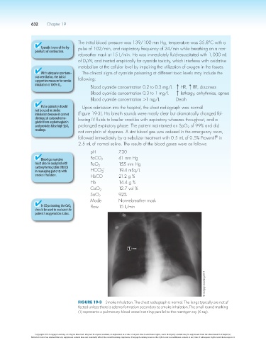
Pulse oximetry should Upon admission into the hospital, the chest radiograph was normal
not be used in smoke
inhalation because it cannot (Figure 19-3). His breath sounds were mostly clear but dramatically changed fol-
distinguish carboxyhemo- lowing IV fluids to basilar crackles with expiratory wheezes throughout, and a
globin from oxyhemoglobin
and provides false high SpO 2 prolonged expiratory phase. The patient maintained an SpO of 99% and did
2
readings. not complain of dyspnea. A stat blood gas was ordered in the emergency room,
®
followed immediately by a nebulizer treatment with 0.5 mL of 0.5% Proventil in
2.5 mL of normal saline. The results of the blood gases were as follows:
pH 7.30
Blood gas samples PaCO 2 41 mm Hg
must also be analyzed with PaO 155 mm Hg
carboxyhemoglobin (HbCO) 2 -
in managing patients with HCO 19.4 mEq/L
3
smoke inhalation. HbCO 21.2 g %
Hb 14.4 g %
CaO 2 12.7 vol %
SaO 2 92%
Mode Non-rebreather mask
In CO poisoning, the CaO 2 Flow 15 L/min
should be used to evaluate the
patient’s oxygenation status.
1
© Cengage Learning 2014
Figure 19-3 Smoke inhalation. The chest radiograph is normal. The lungs typically are not af-
fected unless there is edema formation secondary to smoke inhalation. The small round marking
(1) represents a pulmonary blood vessel running parallel to the roentgen ray (X-ray).
Copyright 2013 Cengage Learning. All Rights Reserved. May not be copied, scanned, or duplicated, in whole or in part. Due to electronic rights, some third party content may be suppressed from the eBook and/or eChapter(s).
Editorial review has deemed that any suppressed content does not materially affect the overall learning experience. Cengage Learning reserves the right to remove additional content at any time if subsequent rights restrictions require it.