
Page 674 - Clinical Application of Mechanical Ventilation
P. 674
640 Chapter 19
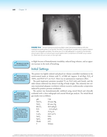
1
2
4
3
© Cengage Learning 2014
Figure 19-4 Tension hemopneumothorax. Right-sided hemopneumothorax shifts the
mediastinum and trachea (1) to the left. The shift is not significant, possibly due to patient rotation
when the radiograph was taken. The white area (2) on the radiograph is caused by the blood in
pleural space. (Note: air in pleural space would appear dark.) Compression of the left lung is noted
(3). Chest tube (4) can be seen on the right side.
to flight because of hemodynamic instability, reduced lung volumes, and an appar-
Hemodynamic instability ent increase in the work of breathing.
is mainly due to blood loss.
Initial Settings
The patient was lightly sedated and placed on volume-controlled ventilation in the
Reduction of lung vol- assist/control mode at 16/min with V of 800 mL (approx. 8 mL/Kg), F O of
umes is caused by compres- T I 2
sion of the lungs by blood and 100%, and PEEP of 5 cm H O. There was no spontaneous respiratory effort.
2
air in the pleural space. The peak inspiratory pressures exceeded 70 cm H O with each breath, and the
2
tidal volume delivered was about 8 mL/kg of body weight. This relatively low vol-
ume maintained adequate ventilation without excessive cardiovascular compromise
induced by positive pressure ventilation.
The patient was hemodynamically stabilized using stored blood and clinically
Increase in work of evaluated with a chest radiograph and arterial blood gas analysis. The initial blood
breathing is caused by
reduction of lung compliance gas results were as follows:
and/or increase of airway
resistance. pH 7.30
PaCO 2 40 mm Hg
PaO 2 83 mm Hg
-
HCO 18.9 mEq/L
3
The peak inspiratory B.E. 26.8 mEq/L
pressure was high (.70 cm Hb 15.8 g %
H 2 O) because of low lung
compliance (due to compres- CaO 2 20.9 vol %
sion of lung parenchyma by
blood and air in the pleural SaO 2 94%
space). SpO 2 89%
Mode A/C
f 16/min
Copyright 2013 Cengage Learning. All Rights Reserved. May not be copied, scanned, or duplicated, in whole or in part. Due to electronic rights, some third party content may be suppressed from the eBook and/or eChapter(s).
Editorial review has deemed that any suppressed content does not materially affect the overall learning experience. Cengage Learning reserves the right to remove additional content at any time if subsequent rights restrictions require it.