
Page 279 - Cardiac Nursing
P. 279
1
1
1
/09
/09
/09
0:2
M
M
Pa
0:2
1 P
1 P
66.
q
q
5-2
5-2
66.
q
6
/29
/29
xd
xd
6
ara
ara
a
p
t
t
a
In
c.
c.
a
a
In
g
g
e 2
Pa
Pa
g
e 2
A
p
p
55
55
A
K34
K34
LWB
0-c
11_
11_
0-c
24
24
LWB
LWBK340-c11_ p p pp245-266.qxd 6/29/09 10:21 PM Page 255 Aptara Inc.
C HAPTER 11 / Laboratory Tests Using Blood 255
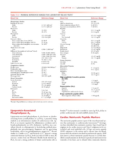
Table 11-4 ■ NORMAL REFERENCE RANGES FOR LABORATORY BLOOD TESTS *
Blood Test Reference Range Blood Test Reference Range
Hematologic Studies SvO 2 60–80%
Red blood cell count Alkaline phosphatase 35–125 IU/L
Males 4.7–6.1 mil/mm 3 Alanine aminotransferase (ALT) 0–40 IU/L
Females 4.2–5.4 mil/mm 3 Aspartate aminotransferase (AST) 5–40 IU/L
Hematocrit Bilirubin
Males 40–50% Total 0.2–1.3 mg/dL
Females 38–47% Direct 0–0.4 mg/dL
Hemoglobin Calcium
Males 13.5–18.0 g/dl Total 8.9–10.3 mg/dL
Females 12.0–16.0 g/dl Free (ionized) 4.6–5.1 mg/dL
Corpuscle indices Creatinine
Mean corpuscular volume (MCV) 82–98 fl Males 0.9–1.4 mg/dL
Mean corpuscular hemoglobin (MCH) 27–31 pg Females 0.8–1.3 mg/dL
Mean corpuscular hemoglobin concentration 32–36% Glucose (fasting) 70–99 mg/dL
(MCHC) LDH 20–200 IU/L
White blood cell count Magnesium 1.3–2.2 mEq/L
Total 4,500–11,000/mm 3 Phosphorus 2.5–4.5 mg/dL
3
Differential (in number of cells/mm blood) Protein (total) 6.5–8.5 g/dL
Total leukocytes 5,000–10,000 (100%) Urea nitrogen 8–26 mg/dL
Total neutrophils 3,000–7,000 (60–70%) Uric acid
Lymphocytes 1,500–3,000 (20–30%) Males 4.0–8.5 mg/dL
Monocytes 375–500 (2–6%) Females 2.8–7.5 mg/dL
Eosinophils 50–400 (1–4%)
Serum Enzymes
Basophils 0–50 (0.1%)
CK-MM 95–100%
Sedimentation rate 0–30 mm/hr
CK-MB 0–5%
Coagulation Studies Myocardial Proteins
Platelet count 250,000–500,000/mm 3
Troponin-I 0–1.6 ng/mL
Prothrombin time 12–15 s
Troponin-T 0–0.1 ng/mL
Partial thromboplastin time 60–70 s
Myoglobin
Activated partial thromboplastin time 35–45 s
Males 20–90 ng/mL
Activated clotting time 75–105 s
Females 10–75 ng/mL
Fibrinogen level 160–300 mg/dL
High sensitivity-C-reactive protein
Blood Chemistries
(hs-CRP)
Serum electrolytes
Low 1.0 mg/L
Sodium 135–145 mEq/L
Average 1.0–3.0 mg/L
Potassium 3.3–4.9 mEq/L
High 3.0 mg/L
Chloride 97–110 mEq/L
Homocysteine (Hcy)
Carbon dioxide 22–31 mEq/L
Optimal 12 μmol/L
Blood gases
Borderline 12–15 μmol/L
pH 7.35–7.45
High Risk for cardiovascular disease 15 μmol/L
35–45 mm Hg
PaCO 2
80–105 mm Hg B-type natriuretic peptide (BNP)
PaO 2
Bicarbonate 22–29 mEq/L Most diagnostic of heart failure 100 pg/mL
Base excess, deficit 0 2.3 mEq/L
*Examples: Regional laboratory techniques and methods may result in variations.
Lipoprotein-Associated females. 68 Further research is needed to assess Lp-PLA 2 ability to
Phospholipase A 2 predict cardiovascular risk and establish plasma levels.
Lipoprotein-associated phospholipase A 2 also known as platelet- Cardiac Natriuretic Peptide Markers
A
activating factor acetylhydrolase or Lp-PLA 2 is presently being
studied as an inflammatory marker of cardiovascular risk. Lp- The natriuretic peptide system is part of the neurohormonal sys-
PLA 2 is an enzyme regulated by inflammatory cytokines and is tem that participates in cardiovascular homeostasis. 69 Investiga-
found predominately on LDL cholesterol. Lp-PLA 2 may con- tors have identified three natriuretic peptides; Type-A and Type-B
tribute directly to atherogenesis by hydrolyzing oxidized phos- originate in the cardiac myocyte and Type-C originates in the en-
pholipids into pro-atherogenic fragments and by generating dothelial and renal epithelial cells. A-Type natriuretic peptide
lysolecithin, which has proinflammatory properties. 65 Recent (ANP) originates in the atrium and is released into the blood-
studies by Caslake et al. 66 and Packard et al. 67 showed a strong stream when the atrium is stretched beyond normal capacity. BNP
A
correlation between Lp-PLA 2 levels and risk of CHD in men. By (also known as brain natriuretic peptide because it was first iden-
contrast, results from the Women’s Health Study showed Lp- tified in small amounts in the brain) is produced and stored in the
PLA 2 was not a strong predictor of future cardiovascular risk in ventricles of the heart and is released when ventricular diastolic
A