
Page 742 - Cardiac Nursing
P. 742
2:0
2:0
1
1
1
4 A
Pa
Pa
M
4 A
M
/09
xd
xd
q
q
q
6
/09
/09
/30
6
/30
Pa
ara
ara
t
p
t
a
c.
c.
In
a
In
p
e 7
e 7
g
g
g
18
A
p
A
18
18
70
70
0-c
K34
K34
29_
29_
29_
p
0-c
5-7
LWBK340-c29_ p pp705-721.qxd 6/30/09 12:04 AM Page 718 Aptara Inc.
5-7
21.
21.
LWB
LWB
718 PA R T I V / Pathophysiology and Management of Heart Disease
Electrocardiography often shows a pattern of left ventricular hy- Medical Management
pertrophy, although its absence does not exclude the presence of
critical aortic stenosis. Patients with aortic stenosis may demon- Because aortic stenosis is a mechanical problem, there is no effec-
strate ST-T-wave changes typical of left ventricular strain. QRS tive medical management. The course of aortic stenosis varies in
voltage changes in the precordial leads correlates poorly with the its progression; therefore, patients should be followed-up carefully
severity of obstruction in aortic stenosis in adults. 10 by their health care providers with serial physical examinations
Exercise testing in patients with mild-to-moderate aortic stenosis and periodic echocardiography. Patients with mild aortic stenosis
with equivocal symptoms may be accomplished with caution in the undergo echocardiography every 2 years. Patients with asympto-
hands of a cardiologist and can provide relevant information regard- matic severe aortic stenosis are followed-up with serial echocar-
ing exercise tolerance. In patients with known severe aortic stenosis diograms every 6 to 12 months and should be taught to recognize
and classic symptoms such as syncope, dyspnea, and chest pain, ex- the symptoms of worsening aortic stenosis, such as dyspnea, chest
ercise testing carries increased risk of ventricular tachyarrhythmias pain, and near syncope or syncope.
and ventricular fibrillation and should not be performed.
Gated blood pool radionuclide scans provide information regard- Interventional and Surgical
ing ventricular function similar to echocardiography and left ven- Management
triculography. Gated pool scans may be useful in patients in
whom left ventriculography cannot be performed (i.e., patients Percutaneous Aortic Catheter Balloon
with elevated creatinine), or those in whom the left ventricle can- Valvuloplasty
not be clearly imaged with echocardiography. Percutaneous aortic catheter balloon valvuloplasty is accom-
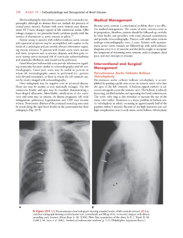
Chest radiography may be negative even in advanced disease. plished by passing a guide wire across the stenotic aortic valve into
Heart size may be normal or only minimally enlarged. The left the apex of the left ventricle. A balloon-tipped catheter is ad-
ventricular border and apex may be rounded, demonstrating a vanced retrograde across the stenotic valve. The balloon is inflated,
boot-shaped silhouette. Identifiable calcification of the aortic fracturing calcified nodules and separating the fused commissures.
valve and aorta may be present. As disease progresses, left atrial The aortic valve ring is also stretched to increase the size of the
enlargement, pulmonary hypertension, and CHF may become aortic valve orifice. Restenosis is a major problem in balloon aor-
evident. Poststenotic dilation of the proximal ascending aorta may tic valvuloplasty in adults, occurring in approximately half of the
be noted along the right heart border in the posteroanterior chest patients within 6 months. Because of the high restenosis rate and
radiograph (Fig. 29-9). high complication rates in early series, aortic balloon valvuloplasty
A B
■ Figure 29-9 (A) Posteroanterior chest radiograph showing rounded border of left ventricle (arrows).s (B) Lat-
eral chest radiograph showing calcified aortic valve (arrowheads) and filling of the retrosternal airspace with dilateds
s
ascending aorta (arrows). (From Boxt, L. M. [1998]. Plain film examination of the chest. In E. J. Topol, R. M.
Califf, J. M. Isner, et al. [Eds.], Textbook of cardiovascular medicine [p. 511]. Philadelphia: Lippincott-Raven.)