
Page 747 - Cardiac Nursing
P. 747
LWBK340-c30_p722-737.qxd 09/09/2009 08:31 AM Page 723 Aptara
C HAP TE R 30 / Pericardial, Myocardial, and Endocardial Disease 723
viral pericarditis is nearly always preceded by a recent respiratory, The pericardium itself does not produce electrical activity. The
gastrointestinal, or “flu-like” illness. A prodrome of fever, malaise, electrocardiogram (ECG) changes seen in pericarditis are a result
and myalgia is common in acute pericarditis, although older pa- of superficial inflammation of the myocardium underneath the
tients may not exhibit fever. 1 pericardium. The ECG of a patient with pericarditis may be nor-
A major symptom of pericarditis is chest pain that is retroster- mal, atypically abnormal with nonspecific changes, or have a four-
nal or left precordial, radiating to the trapezius ridge and varying stage sequence that is diagnostic. Figure 30-1 shows the electro-
with posture. The pain is transmitted through the phrenic nerves, cardiographic manifestations of pericarditis.
and usually occurs on the left side. Shoulder pain should be dis- In stage I, there are ST segment deviations, primarily due to
tinguished from trapezius ridge pain by having the patient physi- inflammation on the ventricular surfaces. PR segment deviations
cally point to the specific site of pain. Frequently the chest pain are also usually present. Stage I is virtually pathognomonic of
caused by pericarditis induces shallow tachypnea as patients at- acute pericarditis when it involves all or almost all leads with early
2
tempt to splint their chest movement. The pain is generally ST junction elevations that produce an appearance of T waves
worse when lying supine and is relieved by sitting. “jacked-up” on the QRS interval, but that is otherwise normal. 2
A pericardial friction rub is pathognomonic for pericarditis, The ST segment is always depressed in aVR. 4
but is frequently not present, may come and go, and can vary in In early stage II, the ST segments return to baseline, and PR
quality and intensity. Auscultation for a pericardial friction rub is segments may now be depressed. In late stage II, the T waves flat-
accomplished with the diaphragm of the stethoscope at the left ten and then invert. In stage III, the ECG is characteristic of dif-
middle to lower sternal border during both inspiration and expi- fuse myocardial injury. In stage IV, the ECG evolves back to the
2
2
ration, while the patient changes positions. Often best heard at prepericarditis state. Stage IV may last days or months. 4
end expiration while the patient is leaning forward, the sound is The changes seen in the ECG of a patient with pericarditis
classically a rasping or creaking with a triple cadence, but can also can occur over hours, particularly from stages I to II, or can take
be bi- or monophasic. 1 place over days or weeks, most often as stage III evolves to stage
A diagnosis of acute pericarditis is made if the patient has a IV. Because of more prompt recognition and treatment of peri-
4
pericardial friction rub or chest pain, and widespread ST segment carditis, not all stages may be exhibited. The ST elevation seen
elevation on electrocardiography. 10 It is important to differenti- in pericarditis is usually distinguished from that of acute MI by
ate pericarditis from myocardial infarction (MI) and pulmonary the absence of Q waves, upward ST segments, and the absence
embolism. Table 30-1 describes the different features from these of associated T wave inversion. 13 In research examining the
three conditions. 10 cause of ST segment abnormalities in emergency department
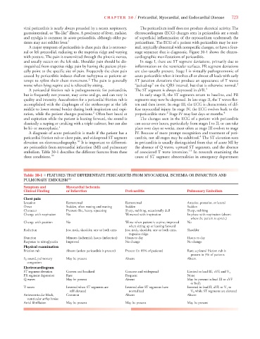
Table 30-1 ■ FEATURES THAT DIFFERENTIATE PERICARDITIS FROM MYOCARDIAL ISCHEMIA OR INFARCTION AND
PULMONARY EMBOLISM 10
Symptom and Myocardial Ischemia
Clinical Finding or Infarction Pericarditis Pulmonary Embolism
Chest pain
Location Retrosternal Retrosternal Anterior, posterior, or lateral
Onset Sudden, often waxing and waning Sudden Sudden
Character Pressure-like, heavy, squeezing Sharp, stabbing, occasionally dull Sharp, stabbing
Change with respiration No Worsened with inspiration In phase with respiration (absent
when the patient is apneic)
Change with position No Worse when patient is supine; improved No
when sitting up or leaning forward
Radiation Jaw, neck, shoulder, one or both arms Jaw, neck, shoulder, one or both arms, Shoulder
trapezius ridge
Duration Minutes (ischemia); hours (infarction) Hours to day Hours to day
Response to nitroglycerin Improved No change No change
Physical examination
Friction rub Absent (unless pericarditis is present) Present (in 85% of patients) Rare; a pleural friction rub is
present in 3% of patients
S 3 sound, pulmonary May be present Absent Absent
congestion
Electrocardiogram
ST segment elevation Convex and localized Concave and widespread Limited to lead III, aVF, and V 1
PR segment depression Rare Frequent None
Q waves May be present Absent May be present in lead III or aVF
or both
T waves Inverted when ST segments are Inverted after ST segments have Inverted in lead II, aVF, or V 1 to
V 4 while ST segments are elevated
still elevated normalized V
Atrioventricular block, Common Absent Absent
ventricular arrhythmias
Atrial fibrillation May be present May be present May be present