
Page 750 - Cardiac Nursing
P. 750
LWBK340-c30_p722-737.qxd 09/09/2009 08:31 AM Page 726 Aptara
726 PA R T IV / Pathophysiology and Management of Heart Disease
acute idiopathic pericarditis. Pericardial effusions occur with Assessment Findings
heart failure and LVH, and are also common after cardiac sur- A 2003 task force of the American College of Cardiology, the Amer-
gery. Effusions associated with cardiac surgery usually resolve af- ican Heart Association, and the American Society of Echocardiogra-
6
ter a month. In a population of patients in Italy, neoplastic eti- phy gave the use of echocardiography for evaluation of all patients
ology was found in 33 of 450 patients with acute pericardial with suspected pericardial disease an evidence class I recommenda-
disease (7.3%). Four percent of these patients presented with tion. 37 Pericardial effusions are classified according to the distance
acute pericardial disease as the first manifestation of their malig- between the left ventricular posterior wall and pericardium. Echocar-
nancy. 35 Pericardial effusions are also seen secondary to uremia diography can classify mild ( 10 mm), moderate (10 to 20 mm),
of renal failure and hypothyroidism. In a population of patients and severe ( 20 mm) effusions. 3,20 “Noncompressing” effusions do
in Turkey, uremic pericarditis resulting from poorly controlled not produce changes in CO or pulsus paradoxus. If the effusions are
renal failure due to economic considerations was the most com- caused by a systemic disease, then the symptoms are related to that
mon cause of pericardial effusion. 20 Tuberculosis is responsible disease. A pericardial rub may or may not be appreciated. The ECG
for approximately 70% of cases of large pericardial effusions in shows reduced voltage, and these changes are nonspecific and unre-
developing countries. 21 liable for diagnosis. Cardiomegaly on chest x-ray film may be ob-
served if effusion is present. If the effusion is visible on radiography,
Pathophysiology then there is at least 250 mL of fluid accumulated. 2
The normal pericardium has a reserve capacity of 150 to 250
mL. An increase of volume of this amount in the pericardial Medical Management
space will not result in a major increase in intrapericardial pres- Pericardial effusion can be treated medically, with pericardiocen-
sure. Intrapericardial pressure will increase once this reserve vol- tesis or with surgery. 20 Patients presenting for the first time with
ume is exceeded, and is also a function of how quickly the vol- pericardial effusion are usually hospitalized to determine the cause
ume in the pericardial space accumulates. If fluid accumulates of the effusion and to observe for the development of cardiac tam-
slowly, the normally stiff pericardium will stretch. However, if ponade. 34 Medical management involves treatment of the peri-
there is increased stiffness of the pericardium, as seen in con- carditis as discussed above with NSAIDs. Conservative treatment
strictive pericarditis, small amounts of fluid will result in in- with clinical and echocardiographic monitoring is usually the ap-
3
creased pericardial pressure. Once the intrapericardial pressure proach for small or moderate effusions. Uremic pericardial effu-
is elevated, the filling of the cardiac chambers becomes limited sions are often treated with aggressive hemodialysis. 20
5
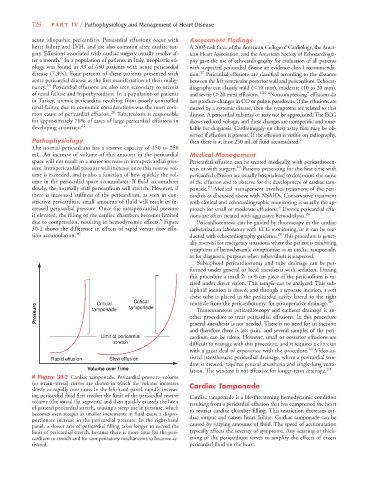
due to compression, resulting in hemodynamic effects. Figure Pericardiocentesis can be guided by fluoroscopy in the cardiac
30-2 shows the difference in effects of rapid versus slow effu- catheterization laboratory with ECG monitoring, or it can be con-
20
sion accumulation. 36 ducted with echocardiography guidance. This procedure is gener-
ally reserved for emergency situations where the patient is exhibiting
symptoms of hemodynamic compromise as in cardiac tamponade,
or for diagnostic purposes when tuberculosis is suspected.
Subxiphoid pericardiostomy and tube drainage can be per-
formed under general or local anesthesia with sedation. During
this procedure a small 2- to 4-cm piece of the pericardium is ex-
cised under direct vision. This sample can be analyzed. This sub-
xiphoid incision is closed, and through a separate incision, a soft
chest tube is placed in the pericardial cavity lateral to the right
Critical
20
ventricle from the pericardiotomy, for postoperative drainage.
Critical
Pressure tamponade tamponade other procedure to treat pericardial effusions. In this procedure
Transcutaneous pericardioscopy and catheter drainage is an-
general anesthesia is not needed. There is no need for an incision
and therefore there is less pain, and several samples of the peri-
Limit of pericardial cardium can be taken. However, small or posterior effusions are
stretch difficult to manage with this procedure, and it requires a clinician
with a great deal of experience with the procedure. 20 Video-as-
Rapid effusion Slow effusion sisted transthoracic pericardial drainage, where a pericardial win-
dow is created, requires general anesthesia and single-lung venti-
Volume over Time 20
lation. The window is not effective for longer-term drainage.
■ Figure 30-2 Cardiac tamponade. Pericardial pressure–volume
(or strain–stress) curves are shown in which the volume increases Cardiac Tamponade
slowly or rapidly over time. In the left-hand panel, rapidly increas-
ing pericardial fluid first reaches the limit of the pericardial reserve Cardiac tamponade is a life-threatening hemodynamic condition
volume (the initial flat segment) and then quickly exceeds the limit resulting from a pericardial effusion that has compressed the heart
of parietal pericardial stretch, causing a steep rise in pressure, which to restrict cardiac chamber filling. This restriction decreases car-
becomes even steeper as smaller increments in fluid cause a dispro-
portionate increase in the pericardial pressure. In the right-hand diac output and causes heart failure. Cardiac tamponade can be
panel, a slower rate of pericardial filling takes longer to exceed the caused by varying amounts of fluid. The speed of accumulation
limit of pericardial stretch, because there is more time for the peri- typically affects the severity of symptoms. Any scarring or thick-
cardium to stretch and for compensatory mechanisms to become ac- ening of the pericardium serves to amplify the effects of excess
tivated. pericardial fluid on the heart.