
Page 62 - untitled
P. 62
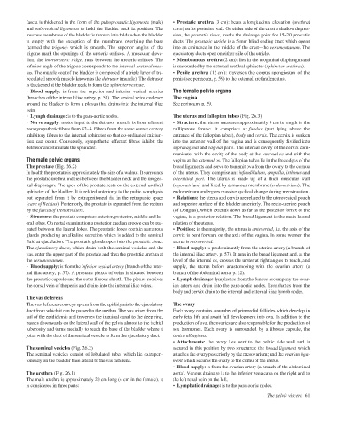
AAAC26  21/5/05  10:45 AM  Page 61
               fascia is thickened in the form of the puboprostatic ligaments (male)
                                                                     crest) on its posterior wall. On either side of the crest a shallow depres-
               and pubovesical ligaments to hold the bladder neck in position. The
                                                                     sion, the prostatic sinus, marks the drainage point for 15–20 prostatic
               mucous membrane of the bladder is thrown into folds when the bladder
                                                                     ducts. The prostatic utricle is a 5 mm blind ending tract which opens
               is empty with the exception of the membrane overlying the base  • Prostatic urethra (3 cm): bears a longitudinal elevation (urethral
               (termed the  trigone) which is smooth. The superior angles of the  into an eminence in the middle of the crestathe verumontanum. The
               trigone mark the openings of the ureteric orifices. A muscular eleva-  ejaculatory ducts open on either side of the utricle.
               tion, the interureteric ridge, runs between the ureteric orifices. The  • Membranous urethra (2 cm): lies in the urogenital diaphragm and
               inferior angle of the trigone corresponds to the internal urethral mea-  is surrounded by the external urethral sphincter (sphincter urethrae).
               tus. The muscle coat of the bladder is composed of a triple layer of tra-  • Penile urethra (15 cm): traverses the corpus spongiosum of the
               beculated smooth muscle known as the detrusor (muscle). The detrusor  penis (see perineum, p. 59) to the external urethral meatus.
               is thickened at the bladder neck to form the sphincter vesicae.
               • Blood supply: is from the superior and inferior vesical arteries  The female pelvic organs
               (branches of the internal iliac artery, p. 57). The vesical veins coalesce  The vagina
               around the bladder to form a plexus that drains into the internal iliac  See perineum, p. 59.
               vein.
               • Lymph drainage: is to the para-aortic nodes.        The uterus and fallopian tubes (Fig. 26.3)
               • Nerve supply: motor input to the detrusor muscle is from efferent  • Structure: the uterus measures approximately 8 cm in length in the
               parasympathetic fibres from S2–4. Fibres from the same source convey  nulliparous female. It comprises a:  fundus  (part lying above the
               inhibitory fibres to the internal sphincter so that co-ordinated micturi-  entrance of the fallopian tubes), body and cervix. The cervix is sunken
               tion can occur. Conversely, sympathetic efferent fibres inhibit the  into the anterior wall of the vagina and is consequently divided into
               detrusor and stimulate the sphincter.                 supravaginal and vaginal parts. The internal cavity of the cervix com-
                                                                     municates with the cavity of the body at the internal os and with the
               The male pelvic organs                                vagina at the external os. The fallopian tubes lie in the free edges of the
               The prostate (Fig. 26.2)                              broad ligaments and serve to transmit ova from the ovary to the cornua
               In health the prostate is approximately the size of a walnut. It surrounds  of the uterus. They comprise an: infundibulum, ampulla, isthmus and
               the prostatic urethra and lies between the bladder neck and the urogen-  interstitial part. The uterus is made up of a thick muscular wall
               ital diaphragm. The apex of the prostate rests on the external urethral  (myometrium) and lined by a mucous membrane (endometrium). The
               sphincter of the bladder. It is related anteriorly to the pubic symphysis  endometrium undergoes massive cyclical change during menstruation.
               but separated from it by extraperitoneal fat in the retropubic space  • Relations: the uterus and cervix are related to the uterovesical pouch
               (cave of Retzius). Posteriorly, the prostate is separated from the rectum  and superior surface of the bladder anteriorly. The recto-uterine pouch
               by the fascia of Denonvilliers.                       (of Douglas), which extends down as far as the posterior fornix of the
               • Structure: the prostate comprises anterior, posterior, middle and lat-  vagina, is a posterior relation. The broad ligament is the main lateral
               eral lobes. On rectal examination a posterior median groove can be pal-  relation of the uterus.
               pated between the lateral lobes. The prostatic lobes contain numerous  • Position: in the majority, the uterus is anteverted, i.e. the axis of the
               glands producing an alkaline secretion which is added to the seminal  cervix is bent forward on the axis of the vagina. In some women the
               fluid at ejaculation. The prostatic glands open into the prostatic sinus.  uterus is retroverted.
               The ejaculatory ducts, which drain both the seminal vesicles and the  • Blood supply: is predominantly from the uterine artery (a branch of
               vas, enter the upper part of the prostate and then the prostatic urethra at  the internal iliac artery, p. 57). It runs in the broad ligament and, at the
               the verumontanum.                                     level of the internal os, crosses the ureter at right angles to reach, and
               • Blood supply: is from the inferior vesical artery (branch of the inter-  supply, the uterus before anastomosing with the ovarian artery (a
               nal iliac artery, p. 57). A prostatic plexus of veins is situated between  branch of the abdominal aorta, p. 32).
               the prostatic capsule and the outer fibrous sheath. The plexus receives  • Lymph drainage: lymphatics from the fundus accompany the ovar-
               the dorsal vein of the penis and drains into the internal iliac veins.  ian artery and drain into the para-aortic nodes. Lymphatics from the
                                                                     body and cervix drain to the internal and external iliac lymph nodes.
               The vas deferens
               The vas deferens conveys sperm from the epididymis to the ejaculatory  The ovary
               duct from which it can be passed to the urethra. The vas arises from the  Each ovary contains a number of primordial follicles which develop in
               tail of the epididymis and traverses the inguinal canal to the deep ring,  early fetal life and await full development into ova. In addition to the
               passes downwards on the lateral wall of the pelvis almost to the ischial  production of ova, the ovaries are also responsible for the production of
               tuberosity and turns medially to reach the base of the bladder where it  sex hormones. Each ovary is surrounded by a fibrous capsule, the
               joins with the duct of the seminal vesicle to form the ejaculatory duct.  tunica albuginea.
                                                                     • Attachments: the ovary lies next to the pelvic side wall and is
               The seminal vesicles (Fig. 26.2)                      secured in this position by two structures: the broad ligament which
               The seminal vesicles consist of lobulated tubes which lie extraperi-  attaches the ovary posteriorly by the mesovarium; and the ovarian liga-
               toneally on the bladder base lateral to the vas deferens.  ment which secures the ovary to the cornu of the uterus.
                                                                     • Blood supply: is from the ovarian artery (a branch of the abdominal
               The urethra (Fig. 26.1)                               aorta). Venous drainage is to the inferior vena cava on the right and to
               The male urethra is approximately 20 cm long (4 cm in the female). It  the left renal vein on the left.
               is considered in three parts:                         • Lymphatic drainage: is to the para-aortic nodes.
                                                                                                        The pelvic viscera 61