
Page 66 - untitled
P. 66
AAAC27 21/5/05 10:45 AM Page 65
Lunate
Triquetral Tubercle of scaphoid
Trapezium
Pisiform
Trapezoid
Capitate
Hook of hamate
Pisiform
Triquetral
Lunate
Flexor retinaculum
Scaphoid
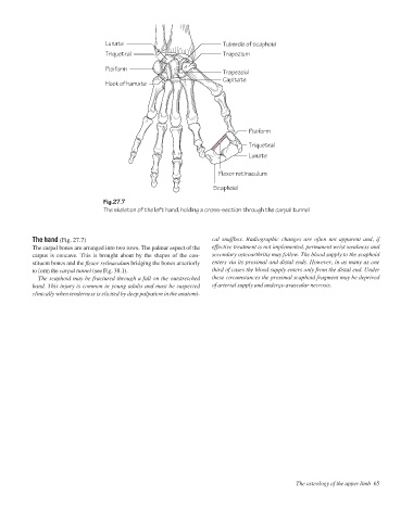
Fig.27.7
The skeleton of the left hand, holding a cross-section through the carpal tunnel
The hand (Fig. 27.7) cal snuffbox. Radiographic changes are often not apparent and, if
The carpal bones are arranged into two rows. The palmar aspect of the effective treatment is not implemented, permanent wrist weakness and
carpus is concave. This is brought about by the shapes of the con- secondary osteoarthritis may follow. The blood supply to the scaphoid
stituent bones and the flexor retinaculum bridging the bones anteriorly enters via its proximal and distal ends. However, in as many as one
to form the carpal tunnel (see Fig. 38.1). third of cases the blood supply enters only from the distal end. Under
The scaphoid may be fractured through a fall on the outstretched these circumstances the proximal scaphoid fragment may be deprived
hand. This injury is common in young adults and must be suspected of arterial supply and undergo avascular necrosis.
clinically when tenderness is elicited by deep palpation in the anatomi-
The osteology of the upper limb 65