
Page 142 - Color Atlas Of Pathophysiology (S Silbernagl Et Al, Thieme 2000)
P. 142
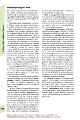
Pathophysiology of Bone
       Bone consists of connective tissue or bone ma-  However, most often the cause remains un-
       trix (sulfate-containing proteoglycans, glyco-  known (primary osteoporosis).
       proteins and hydroxyproline-containing col-  Effects of osteoporosis include skeletal pain
       lagen fibers) and the bone minerals (alkaline  even at rest, intervertebral disc prolapse, lower
                            2–
                                2+
              2+
                         +
       salts of Ca , phosphate, Na , CO 3 , Mg , K , +  arm or femoral neck fractures. Hypercalcemia
           –
       and F ).                        may be present in extreme cases. Depending
         Construction and mineralization. Formation  on its cause, the osteoporosis may be localized
       of the bone matrix is promoted, among others,  (e.g., under a rigid cast) or generalized (e.g.,
    Kidney, Salt and Water Balance  It is probably initiated through splitting of py-  growth plate is disturbed (→ A5). Before longi-
                                       due to excess glucocorticoids).
       by insulin and inhibited by glucocorticoids.
                                        In osteomalacia and rickets the mineraliza-
         Bone mineralization is inhibited by pyro-
                                       tion of the bone matrix (osteoid) or of the
       phosphate (two esterified phosphoric acids).
       rophosphate by alkaline phosphatase. The plas-
                                       tudinal growth is concluded and before epiph-
       ma concentration of this enzyme, produced by
                                       yseal fusion has occurred, the abnormality
       osteoblasts, is a measure of osteoblast activity.
                                       mostly leads to rickets (widening of the growth
                                       plates and distorted growth). After longitudi-
       The mineralization is fostered by increase of
         2+
                                       nal growth has ceased the decreased minerali-
          and phosphate plasma concentration—an
       Ca
                  occurs
                                       the course of normal bone remodeling), leads
                        in
       (1,25-[OH] 2 -D 3 )
                                 steps
                           several
       (→ A1): under the influence of ultraviolet light
                                       to osteomalacia. Both rickets and osteomalacia
       vitamin D 3 is formed in the skin from 7-dehy-
                                       can be caused by a reduced formation of calci-
    5  effect of calcitriol. The formation of calcitriol  zation of the newly formed osteoid (formed in
       drocholesterol. Vitamin D 3 is converted in the  triol, for example, in lack of ultraviolet light
       liver to 25-OH 2 -D 3 under the influence of es-  and of vitamin D, by estrogen deficiency (post-
       trogens, and in the kidney to 1,25-(OH) 2 -D 3 un-  menopausal), or by renal failure (→ p.110ff.).
       der the influence of PTH. The building up of  Even without calcitriol deficiency, hypophos-
       bone and mineralization are additionally stim-  phatemia (phosphate diabetes, Fanconi’s syn-
       ulated by mechanical use of the bone.  drome; → p. 96, 110ff.) or chronic renal tubular
         The breaking down of the bone matrix leads  acidosis can result in osteomalacia. Osteoma-
       to the increased renal excretion of hydroxypro-  lacia can occur in dialyzed patients who suffer
       line (→ A2), while demineralization leads to  from aluminum intoxication. Lastly, a rickets-
       increased renal excretion of Ca 2+  and phos-  like or osteomalacia-like clinical syndrome oc-
       phate (urolithiasis; → A3).     curs in the rare, genetic deficiency of alkaline
         The breakdown of bone is, among other fac-  phosphatase (hypophosphatasia).
       tors, due to lacking mechanical stress (immo-  The effects of rickets are retarded growth,
       bilization). Localized breakdown of bone can  bow-legs or knock-knees, vertebral column
       be caused by osteoclast activating factor  deformities, prominence of the costochondral
       (OAF), which in tumors results in demineral-  junctions (rachitic rosary) as well as thin and
       ization of bone.                soft cranial, particularly occipital, bones (cra-
         The most important abnormalities of bone  niotabes). Osteomalacia leads to bone pain
       are osteopenia (or osteoporosis) and osteoma-  (pain on movement), translucent bands of de-
       lacia (or rickets in children). Osteopenia is de-  mineralization in bone (pseudofractures or
       fined as reduction of bone mass below the  Looser’s zones), and muscular weakness (Ca 2+
       norm for age, race, and sex, caused by prolong-  deficiency).
       ed imbalance between buildup and break-
       down of bone. Osteoporosis is defined as the
       clinical state resulting from reduced bone
       mass (→ A4). Causes include excess glucocor-
       ticoids, lack of estrogen (postmenopausal), in-
  132  sulin deficiency (diabetes mellitus), and inac-
       tivity (rigid cast, tetraplegia, microgravity).
       Silbernagl/Lang, Color Atlas of Pathophysiology © 2000 Thieme
       All rights reserved. Usage subject to terms and conditions of license.