
Page 170 - Color Atlas Of Pathophysiology (S Silbernagl Et Al, Thieme 2000)
P. 170
Chronic Pancreatitis
Chronic pancreatitis is an inflammatory pro- proenzyme content (while the concentration
cess that destroys the exocrine and endo- of trypsin inhibitor–protein remains un-
crine tissues and leads to fibrosis of the or- changed; → p.158).
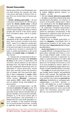
gan. There are several forms of chronic pan- ! The rarer chronic–obstructive pancreatitis
creatitis: (→ A, right) is caused by occlusion of the main
! Chronic calcifying pancreatitis (→ A, left) excretory duct(s) by tumors, scar stricture, or
is the most common form (70–80% of cases), stenosis of the papilla, among others. There
caused by chronic alcohol abuse (> 80 g/d is no calcification, but the ductal system is
over many years) and is characterized by ir- markedly dilated upstream of the stenosis
regularly distributed tissue lesions with in- (→ A; endoscopic retrograde pancreatogra-
Liver traductal protein plugs and stones as well as phy [ERP], in which contrast media are in-
atrophy and stenosis of the ductal system.
jected for radiological visualization). If the
Stomach, Intestines, genesis: chronic pancreatitis (in contrast to the calci-
obstruction is removed in time, this form of
Three mechanisms play a role in its patho-
1. While normally, in parallel with the
fying form) is reversible.
stimulation of the acini (enzyme-rich secre-
! Other forms of chronic pancreatitis in-
tion), there is greater secretion in the ducts
clude the idiopathic, nonalcoholic form in
–
malnourished juveniles in the tropics, and
(HCO 3 , water), it is reduced in chronic pan-
in the pancreatic juice is increased, especial-
parathyroidism.
Acute exacerbation of chronic pancreatitis
ly when acinar secretion is stimulated. This
6 creatitis. As a result, protein concentration the form seen in hypercalcemia due to hyper-
leads to protein precipitation in the ductal is usually difficult to distinguish from acute
lumens and protein plugs and deposits are pancreatitis, especially when there is a his-
thus formed. tory of high alcoholic intake. In both cases
2. Calcium salts are deposited on the pre- the premature activation of pancreatic en-
cipitated protein, resulting in the formation zymes is a prominent feature (see above and
of stones in the lumen of small ducts, and p.158). It can lead, via pancreatic edema, to
concentric calcium deposits on the walls of pancreatic hemorrhage and necrosis as well
the larger ducts. The cause of all this may be as to acute pseudocysts, abscess, and/or im-
that two components of pancreatic juice are pairment of neighboring organs such as duo-
diminished in chronic pancreatitis, namely denum, antrum, choledochal duct, and colon.
those that normally prevent the precipita- The results of chronic pancreatitis are tis-
tion of calcium salts from pancreatic juice. sue atrophy, ductal stenosis, and periductal
One of these components is citrate, which fibrosis with scarring. This gradually leads
binds calcium complexly, the other is the to loss of parenchyma, which will cause exo-
14 kDa protein, lithostatin (= pancreatic crine and later also endocrine pancreatic in-
stone protein [PSP]), which holds calcium sufficiency. Intermittent or continuous pain,
salts in solution during (physiological) hy- malabsorption (→ p.152ff.), diarrhea (→
persaturation. p.150), and weight loss as well as diabetes
3. Similar to acute pancreatitis (→ p.158), mellitus (→ p. 286ff.) and damage to neigh-
intraductal activation of trypsin occurs. This boring organs (pancreatic ascites, portal and
not only contributes to the autodigestion of splenic vein thrombosis, obstructive jaun-
pancreatic tissue, but also activates other ag- dice, etc.) are associated with this.
gressive enzymes, such as elastase and phos-
pholipase A 2 , in the ductal system and, in
some circumstances, also interstitially. It is
thought that the cause of the premature en-
zyme activation is that impaired drainage
160 has increased intraductal pressure, resulting
in epithelial lesions, together with raised
Silbernagl/Lang, Color Atlas of Pathophysiology © 2000 Thieme
All rights reserved. Usage subject to terms and conditions of license.