
Page 349 - ACCCN's Critical Care Nursing
P. 349
326 P R I N C I P L E S A N D P R A C T I C E O F C R I T I C A L C A R E
Thyroid cartilage bronchi. Further divisions within these conducting
Cricoid cartilage airways end with the terminal bronchioles, the smallest
Trachea Upper lobe airways without alveoli. These conducting airways do not
Clavicle of left lung
Upper lobe participate in gas exchange but form the anatomical dead
7
of right lung space (approximately 150 mL).
Scapula 1 1 Larger airways have a greater proportion of supporting
cartilage, ciliated epithelium, goblet and serous cells and
2 2 hence a mucous layer. As the airways become smaller,
cartilage becomes irregularly dispersed, the number of
3 3
goblet cells and amount of mucus decreases until, at the
Sternum 4 4 alveolar level, there is only a single layer of squamous
epithelial cells. Alveolar macrophages are present in these
5 5 epithelial cells, and phagocytose any small particles that
6 6 may enter the alveolar area. Smooth muscle surrounds
Lower lobe and supports the bronchioles, enabling airway diameter
Middle lobe 7 7 of left lung change and subsequent changes in airway resistance to
Lower lobe 8 8 gas flow. 8
9 9
Rib cartilages
10 10 THORAX/LUNGS
The lungs and heart are protected within the thoracic
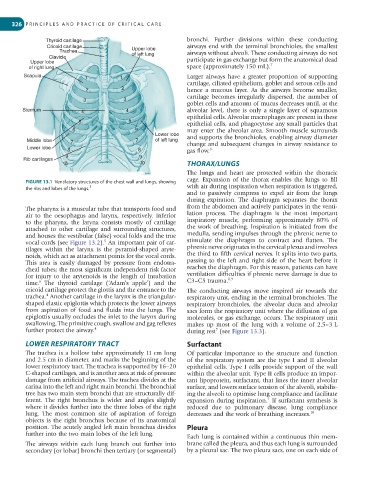
FIGURE 13.1 Ventilatory structures of the chest wall and lungs, showing cage. Expansion of the thorax enables the lungs to fill
1
the ribs and lobes of the lungs. with air during inspiration when respiration is triggered,
and to passively compress to expel air from the lungs
during expiration. The diaphragm separates the thorax
The pharynx is a muscular tube that transports food and from the abdomen and actively participates in the venti-
air to the oesophagus and larynx, respectively. Inferior lation process. The diaphragm is the most important
to the pharynx, the larynx consists mostly of cartilage inspiratory muscle, performing approximately 80% of
attached to other cartilage and surrounding structures, the work of breathing. Inspiration is initiated from the
and houses the vestibular (false) vocal folds and the true medulla, sending impulses through the phrenic nerve to
5
vocal cords (see Figure 13.2). An important pair of car- stimulate the diaphragm to contract and flatten. The
tilages within the larynx is the pyramid-shaped aryte- phrenic nerve originates in the cervical plexus and involves
noids, which act as attachment points for the vocal cords. the third to fifth cervical nerves. It splits into two parts,
This area is easily damaged by pressure from endotra- passing to the left and right side of the heart before it
cheal tubes; the most significant independent risk factor reaches the diaphragm. For this reason, patients can have
for injury to the arytenoids is the length of intubation ventilation difficulties if phrenic nerve damage is due to
8,9
6
time. The thyroid cartilage (‘Adam’s apple’) and the C3–C5 trauma.
cricoid cartilage protect the glottis and the entrance to the The conducting airways move inspired air towards the
4
trachea. Another cartilage in the larynx is the triangular- respiratory unit, ending in the terminal bronchioles. The
shaped elastic epiglottis which protects the lower airways respiratory bronchioles, the alveolar ducts and alveolar
from aspiration of food and fluids into the lungs. The sacs form the respiratory unit where the diffusion of gas
epiglottis usually occludes the inlet to the larynx during molecules, or gas exchange, occurs. The respiratory unit
swallowing. The primitive cough, swallow and gag reflexes makes up most of the lung with a volume of 2.5–3 L
further protect the airway. 4 during rest (see Figure 13.3).
7
LOWER RESPIRATORY TRACT Surfactant
The trachea is a hollow tube approximately 11 cm long Of particular importance to the structure and function
and 2.5 cm in diameter, and marks the beginning of the of the respiratory system are the type I and II alveolar
lower respiratory tract. The trachea is supported by 16–20 epithelial cells. Type I cells provide support of the wall
C-shaped cartilages, and is another area at risk of pressure within the alveolar unit. Type II cells produce an impor-
damage from artificial airways. The trachea divides at the tant lipoprotein, surfactant, that lines the inner alveolar
carina into the left and right main bronchi. The bronchial surface, and lowers surface tension of the alveoli, stabilis-
tree has two main stem bronchi that are structurally dif- ing the alveoli to optimise lung compliance and facilitate
ferent. The right bronchus is wider and angles slightly expansion during inspiration. If surfactant synthesis is
7
where it divides further into the three lobes of the right reduced due to pulmonary disease, lung compliance
lung. The most common site of aspiration of foreign decreases and the work of breathing increases. 10
objects is the right bronchus because of its anatomical
position. The acutely angled left main bronchus divides Pleura
further into the two main lobes of the left lung. Each lung is contained within a continuous thin mem-
The airways within each lung branch out further into brane called the pleura, and thus each lung is surrounded
secondary (or lobar) bronchi then tertiary (or segmental) by a pleural sac. The two pleura sacs, one on each side of