
Page 351 - ACCCN's Critical Care Nursing
P. 351
328 P R I N C I P L E S A N D P R A C T I C E O F C R I T I C A L C A R E
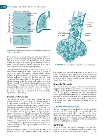
Inspiration Expiration
Intrapulmonary
pressure
+2
0
Pressure relative to atmospheric pressure (mm Hg) −2 Trans- Pulmonary
pulmonary
vein to
pressure
−4
left heart
−6
Intrapleural
pressure
Pulmonary
−8
artery from
plexus
right heart Capillary
Volume of breath
Volume (L) 0 Alveoli
0.5
5 seconds elapsed
FIGURE 13.4 Changes in intrapleural and intrapulmonary pressure during
4
inspiration and expiration.
the midline, are completely separate from each other. The
parietal pleura lines the inner surface of the chest wall
and is in close contact with the visceral pleura, which
covers the lungs. The pleural space, between these two
layers, contains a small amount of serous fluid, which FIGURE 13.5 Terminal ventilation and perfusion units of the lung. 5
normally limits friction during lung expansion.
The intra-pleural pressure in the pleural space under
normal circumstances is always negative with a range of
−4 to −10 cmH 2 O; this negative pressure keeps the lungs remarkably low (normal pulmonary artery pressure is
7
inflated. During inhalation the pressure becomes more only 25/8 mmHg; mean 15 mmHg). This low pressure
negative as both the lungs and the chest wall are elastic system ensures that the work of the right heart is as small
structures. These elastic fibres of the lung pull the visceral as feasible, while promoting efficient gas exchange in the
11
pleura inwards while the chest wall pulls the parietal lungs (see Figure 13.6).
pleura outward. The pressure difference between the alve-
olar pressure (0 cmH 2 O pressure in the lungs) and the Bronchial Circulation
intra-pleural pressure (−4 cmH 2 O) across the lung wall The bronchial circulation, part of the systemic circulation,
is termed the trans-pulmonary pressure (+4 cmH 2 O supplies oxygenated blood, nutrients and heat to the con-
[0 − (−4) = +4]), and is the force that hold the lungs ducting airways (to the level of the terminal bronchioles)
open (see Figure 13.4). and to the pleura. Drainage of this deoxygenated
3,4
blood is predominantly through the bronchial network,
Pulmonary Circulation although some capillaries drain into the pulmonary arte-
The circulatory system of the lung receives the entire rial circulation, contributing to venous admixture or
7
cardiac output but operates as a low pressure system, as right-to-left shunt (see Pathophysiology below for further
it only directs blood back to the left side of the heart discussion).
(unlike the systemic circulation which pumps blood to
different regions of the entire body). The pulmonary cir- CONTROL OF VENTILATION
culation involves oxygen-depleted blood being pumped Normal breathing occurs automatically and is a complex
by the right ventricle to the lungs via the pulmonary function not fully understood. It is coordinated by the
artery, with oxygen-rich blood returning to the left atrium respiratory centre, regulated by controllers in the brain,
via the pulmonary veins. Pulmonary blood vessels follow effectors in the muscles and sensors including chemore-
the path of the bronchioles, with the capillaries forming ceptors and mechanoreceptors. There are also protective
a dense network in the walls of the alveoli. As illustrated reflexes that respond to irritation of the respiratory tract
5
in Figure 13.5, the entire surface area of the alveolar wall such as coughing and sneezing.
is covered by these capillaries, where gas exchange occurs
as the capillaries are just large enough for a red blood cell Controller
to pass through.
In the brainstem, the medulla oblongata and the pons
Pulmonary vessels are short, thin and have relatively little regulate automatic ventilation while the cerebral cortex
smooth muscle. The pressure inside the vessels is regulates voluntary ventilation (see Figure 13.7). The