
Page 352 - ACCCN's Critical Care Nursing
P. 352
Respiratory Assessment and Monitoring 329
respiratory rhythmic centre in the medulla can be divided Effectors
into inspiratory and expiratory centres, with the follow- The diaphragm is the major muscle of inspiration, although
ing functions: 8
the external intercostal muscles are also involved. The
● The inspiratory centre (or dorsal respiratory group) accessory muscles of inspiration (scalenes, sternocleido-
triggers inspiration. masteoid muscles and the pectoralis minor of the thorax)
● The expiratory centre (or ventral respiratory group) are active only during exercise or strenuous breathing.
only functions during forced respiration and active Expiration is a passive act and only the internal intercostal
expiration. muscles are involved at rest. During exercise, the abdomi-
4
● The pneumotaxic and apneustic centre in the pons nal muscles also contribute to expiration. Inspiration is
adjusts the rate and pattern of breathing. triggered by stimulus from the medulla, causing the dia-
● The cerebral cortex provides conscious voluntary phragm to contract downwards, and the external intercos-
control over the respiratory muscles. This voluntary tal muscles to contract, lifting the thorax up and out. This
control cannot be maintained when PCO 2 and hydro- action lowers pressure within the alveoli (intra-alveolar
gen ion (H ) concentration become markedly ele- pressure) relative to atmospheric pressure. Air rushes into
+
vated; an example is the inability to hold your breath the lungs to equalise the pressure gradient. After contrac-
8
for very long. Emotional and autonomic activities tion has ceased, the ribs and diaphragm relax, the pressure
also often affect the pace and depth of breathing. gradient reverses, and air is passively expelled from the
lungs and return to their resting state due to elastic recoil.
Mean = 15 Mean = 100
Sensors
25 / 8 120 / 80
Artery Artery
A chemoreceptor is a sensor that responds to a change in
12 Pulmonary Systemic 30 the chemical composition of the blood; there are two
types: central and peripheral. Central chemoreceptors
25 / 0 120 / 0
account for 70% of the feedback controlling ventilation,
Cap RV LV Cap 20 and respond quickly to changes in the pH of cerebral
9
spinal fluid (CSF) (increase of PCO 2 in arterial blood).
RA LA If the PCO 2 in arterial blood remains high for a pro-
8 2 5 longed period, as in chronic obstructive pulmonary
10 disease (COPD), a compensatory change in HCO 3 occurs
and the pH in CSF returns to its near normal value.
7
Vein Vein Under these conditions a patient breathes due to hypoxic
drive; that is, low levels of O 2 are detected by peripheral
FIGURE 13.6 Comparison of pressure in the pulmonary and systemic cir- chemoreceptors and this triggers breathing. For this small
7
culations (mmHg). percentage of the population with COPD, care is required
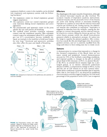
Higher brain centers
(cerebral cortex-voluntary
control over breathing)
Other receptors (e.g. pain)
and emotional stimuli acting ±
through the hypothalamus
±
Respiratory centers
(medulla and pons)
Peripheral
chemoreceptors +
+
O 2 ↓,CO 2 ↑,H ↑ Stretch receptors
in lungs
+ −
Central
chemoreceptors
+
CO 2 ↑,H ↑ −
+ Irritant
FIGURE 13.7 Respiratory centres and reflex receptors
4
controls. (Elaine N. Marieb and Katja Hoehn, Receptors in
HUMAN ANATOMY & PHYSIOLOGY, 8th Ed. © muscles and joints
2010, p. 836. Reprinted by permission of
Pearson Education, Inc., Upper Saddle River,
New Jersey).