
Page 669 - ACCCN's Critical Care Nursing
P. 669
646 S P E C I A LT Y P R A C T I C E I N C R I T I C A L C A R E
in a closed spaced as well as if there are facial burns,
singed nasal hairs or carbonaceous debris in the mouth
71
or pharynx or in the sputum. The specific changes are 9%
dependent on the type of substances inhaled at the time
of injury. In addition, the size of the smoke particles that
are inhaled will affect the location of any injury. If coarse
smoke particles are inhaled, these will primarily be
deposited in the upper tracheobronchial tree, whilst fine 18% 18%
smoke particles will usually be lodged in the alveoli.
Patients with inhalation burn injury will usually experi- Front
ence upper airway oedema and bronchospasm in the 9% 18% 9%
early stages, with the airway disease progressing to the 18%
small airways in subsequent days. 71,72,75 Back 9% Front
1% 9%
Clinical Manifestations 18%
The most prominent clinical manifestations of burn Back
injury are the dermal signs of injury. ANZBA categorise
burns as follows: 74 18% 18%
1. Epidermal burns are limited to injury to the epi-
dermis and tend to be very painful, with a common 14% 14%
example being sunburn. The skin is pink to red in
colour and remains intact. The surrounding tissues
may be oedematous and there is no blistering. This
burn injury will usually heal within 7 days.
2. Superficial partial-thickness burn injury involve A B
the epidermal and superficial dermal layers and are 78
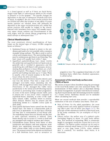
generally red or mottled in appearance and the FIGURE 23.7 Diagram of the ‘rule of nines’ (A), adult; (B), child.
underlying skin will blanch with pressure, demon-
strating that perfusion is intact; blisters are a hall-
mark symptom. This degree of burn injury is very
painful and healing may take up to 14 days. There pinprick is lost. The coagulated dead skin of a full
is usually a lot of wound exudate in the first 72 thickness burn, which has a leathery appearance,
hours where the skin is broken. is called eschar.
3. Mid-dermal partial-thickness injuries extend a part
way into the dermis. They have a large zone of Assessment of the total body surface area
damaged non-viable tissue extending into the (TBSA) of burns
dermis, with damaged but viable tissue at the base. The extent of injury is best described using the percentage
Preservation of the damaged but viable tissue of the total body surface area that sustained burns. The
(particularly in the initial period following injury) measurement of burn surface area is important during
is pivotal to preventing burn wound progression. the initial management of people with burns for estimat-
As some of the nerve endings remain viable, pain ing fluid requirements and determining need for transfer
is present but is less severe when compared to to a burns service. Erythema should not be included
superficial burns. Similarly, as some of the capil- when calculating burn area.
laries remain viable, capillary return is present, There are several methods that provide a reproducible
albeit delayed. Blisters may be present and the estimation of the area of surface area burns. These are:
underlying dermis is a variable colour (pale to
dark pink). l Rule of Nines: for the adult population, the most
4. Deep partial-thickness burns extend into the deep widely known and easily applied method of estimat-
dermal layer. The tissue is a characteristic pink to ing TBSA is the ‘rule of nines’ (see Figure 23.7). The
pale ivory in appearance. It can also have a blotchy principle of this assessment method is that most areas
red base due to extravasation of red blood cells. of the body constitute 9% (or multiples of 9%) of
The underlying tissue does not blanch and the hair the TBSA.
is easily removed; sensation is reduced. These l Palmar surface: the surface area of a patient’s palm
burns usually take in excess of 3 weeks to heal and (including fingers) is about 1% total body surface
are managed with surgical excision and closure. area. This method of estimating TBSA is commonly
5. Full-thickness burns destroy both layers of skin taught in emergency medicine courses but is yet to be
(epidermis and dermis) and may penetrate more validated. The Palmar surface method can be used to
deeply into underlying structures. These burns estimate relatively small burns (<15% of total surface
have a dense white, waxy or even charred appear- area) or very large burns (>85%, when unburnt
ance. The sensory nerves in the dermis are destroyed skin is counted). For medium sized burns, it is
in a full thickness burn, and so sensation to inaccurate.