


Page 173 - Concise Pathology for Exam Preparation ( PDFDrive )
P. 173
158   SECTION I  General Pathology

                                                               First exposure
                                   Antigen-                      to antigen
                                  presenting
                                       cell
                                     (APC)                        Antigen
                                                                (Tubercular
                                 Antigen receptor  MHC            protein)
                                               class II

                                                IL–I2


                                                                Naive
                                                                CD4   T cell
                                              Antigen
                                     APC
                                 (Macrophage/                 Differentiation
                                 dendritic cell)
                                                                TH1 cell

                                                              γ Interferon


                                                 Macrophage       Activated
                                                                 macrophage


                                  Killing of mycobacteria  • Formation of phagolysosome
                                      (immunity)          • ↑ No
                                                          • ↑Generation of free radicals


                                  Monocyte recruitment    • ↑ Tumour necrosis factor (TNF)


                                Differentiation of monocytes  • ↑ Expression of MHC class II
                                  into epithelioid cells   molecules and increased antigen
                                                           presentation
                                                          • ↑ IL–1 and IL–I2
                                    Granulomatous         • ↑ PDGF       ↑Collagen
                                response (hypersensitivity)              synthesis
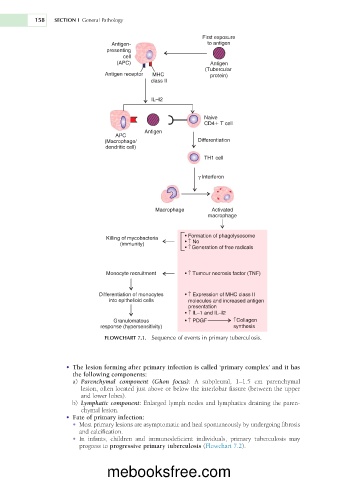
                                 FLOWCHART 7.1.  Sequence of events in primary tuberculosis.


                     •  The lesion forming after primary infection is called ‘primary complex’ and it has
                       the following components:
                        a)  Parenchymal  component  (Ghon  focus):  A  subpleural,  1–1.5  cm  parenchymal
                          lesion, often located just above or below the interlobar fissure (between the upper
                          and lower lobes).
                        b)  Lymphatic component: Enlarged lymph nodes and lymphatics draining the paren-
                          chymal lesion.
                     •  Fate of primary infection:
                       •  Most primary lesions are asymptomatic and heal spontaneously by undergoing fibrosis
                         and calcification.
                       •  In  infants,  children  and  immunodeficient  individuals,  primary  tuberculosis  may
                         progress to progressive primary tuberculosis (Flowchart 7.2).


                                  mebooksfree.com

