


Page 177 - Concise Pathology for Exam Preparation ( PDFDrive )
P. 177
162   SECTION I  General Pathology


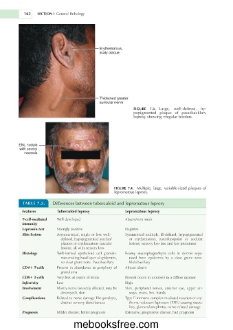
                                              Erythematous,
                                              scaly plaque


                                              Thickened greater
                                              auricular nerve
                                                             FIGURE  7.3.  Large,  well-defined,  hy-
                                                             popigmented  plaque  of  paucibacillary
                                                             leprosy showing irregular borders.


         ENL nodule
          with central
            necrosis


                                                    FIGURE  7.4.  Multiple,  large,  variable-sized  plaques  of
                                                    lepromatous leprosy.

           TABLE 7.3.    Differences between tuberculoid and lepromatous leprosy
           Features        Tuberculoid leprosy           Lepromatous leprosy

           T-cell-mediated    Well developed             Absent/very weak
           immunity
           Lepromin test   Strongly positive             Negative
           Skin lesions    Asymmetrical, single or few, well-   Symmetrical multiple, ill-defined, hypopigmented
                            defined, hypopigmented patches/  or  erythematous,  maculopapular  or  nodular
                            plaques or erythematous macular   lesions; sensory loss late and less prominent
                            lesions; all with sensory loss
           Histology       Well-formed  epithelioid  cell  granulo-  Foamy  macrophages/lepra  cells  in  dermis  sepa-
                            mas eroding basal layer of epidermis,   rated  from  epidermis  by  a  clear  grenz  zone.
                            no clear grenz zone. Paucibacillary  Multibacillary
           CD41 T-cells    Present  in  abundance  at  periphery  of   Almost absent
                            granuloma
           CD81 T-cells    Very few; at centre of lesion   Present (more in number) in a diffuse manner
           Infectivity     Low                           High
           Involvement     Mostly nerve (severely affected, may be   Skin,  peripheral  nerves,  anterior  eye,  upper  air-
                            destroyed), skin               ways, testes, feet, hands
           Complications   Related to nerve damage like paralysis,   Type II immune complex-mediated reaction or ery-
                            distinct sensory disturbances  thema nodosum leprosum (ENL) causing vascu-
                                                           litis, glomerulonephritis, nerve-related damage
           Prognosis       Milder disease; better prognosis  Extensive, progressive disease; bad prognosis

                                  mebooksfree.com

