


Page 175 - Concise Pathology for Exam Preparation ( PDFDrive )
P. 175
160   SECTION I  General Pathology


                         drain through lymphatics into lymphatic ducts, which empty into the right side of
                         heart and then into pulmonary arteries. When the dissemination involves only lungs,
                         it is called miliary pulmonary tuberculosis. Systemic miliary tuberculosis ensues
                         when infective foci in lungs seed pulmonary venous return to the heart; the organ-
                         isms subsequently disseminate through systemic arterial system. Almost every organ
                         in body can be seeded.
                       •  Isolated-organ tuberculosis is a consequence of haematogenous seeding and organs
                         that are typically involved include meninges (tuberculous meningitis), genital or-
                         gans and urinary tract (genitourinary tuberculosis), adrenals (formerly an impor-
                         tant  cause  of  Addison  disease)  and  bones  (osteomyelitis).  When  vertebrae  are
                         affected, the disease is referred to as Pott disease. Paraspinal ‘cold abscesses’ in these
                         patients may track along tissue planes to present as an abdominal or pelvic mass. The
                         most common type of extra pulmonary tuberculosis is lymphadenitis and it most
                         frequently involves the cervical region. The usual presentation is that of discharging
                         sinuses with an underlying cervical swelling (‘scrofula’). The lymph nodes are in-
                         volved as a consequence of lymphatic spread.

                     Microscopy
                     Epithelioid cell granulomas with or without caseation are the histological hallmark of tu-
                     berculous disease. These granulomas are usually enclosed within a fibroblastic rim. Multi-
                     nucleate giant cells called ‘Langhans giant cells’ are present in the granuloma along with
                     mononuclear cells including lymphocytes, plasma cells and histiocytes. Immunocompro-
                     mised individuals do not form well-defined granulomas and may manifest with ill-formed
                     aggregates of histiocytes and chronic inflammatory cells (see Chapter 2).
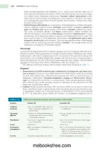
                        The differences between primary and secondary tuberculosis are listed in Table 7.2.
                     Diagnosis
                       1.  Demonstration of AFB on microscopic examination of a diagnostic specimen (spu-
                        tum or tissue): Smears or tissue slides stained with Ziehl Neelsen stain are examined
                        for AFB. This method has a relatively low sensitivity (40–60%) in confirmed cases of
                        pulmonary  tuberculosis.  Auramine-rhodamine  staining  and  fluorescence  micros-
                        copy can improve the sensitivity to a certain extent. Three sputum specimens, prefer-
                        ably collected early in the morning, should be submitted to the laboratory for AFB
                        smear and mycobacterial culture.
                       2.  Culture: Besides sputum and tissue, other specimens which can be used for culture are
                        body cavity fluids, urine or gastric lavage fluid. Specimens may be inoculated onto egg-
                        or agar-based medium (eg, Löwenstein–Jensen or Middlebrook 7H10) and incubated
                        at 37°C. M. tuberculosis grows slowly (4–8 weeks). A presumptive diagnosis can be

           TABLE 7.2.    Differences between primary and secondary tuberculosis

           Features          Primary TB                      Secondary TB
           Evolution         Seen  in  individuals  who  have  not   Occurs due to reactivation of a primary focus
           of disease         been  previously  sensitized  to  tu-  or reinfection
                              bercle bacilli
           Age group         Common  in  children/individuals  of   Any  age  (usually  occurs  later  than  primary
           affected           younger age; may be seen in adults   infection)
                              in developed countries
           Distribution      Lower part of upper lobe and upper   Apex of one or both lobes due to high oxygen
                              part of lower lobe               tension in apices
           Lesion            Ghon focus                      Simon focus
           Involvement       Early involvement of lymphatics and   Due  to  pre-existing  hypersensitivity,  bacilli
           of lymphatics      lymph nodes                      induce an immediate tissue response that
                                                               walls off the lesion and prevents early in-
                                                               volvement of lymphatics and lymph nodes
           Severity          Generally asymptomatic, less severe  Usually symptomatic, more severe


                                  mebooksfree.com

