


Page 174 - Concise Pathology for Exam Preparation ( PDFDrive )
P. 174
7  Infections  159


                               Pneumonia-like consolidation of middle and lower lobes,

                             hilar lymphadenopathy and pleural effusion (cavitation is rare)
                                   Lymphatic or haematogenous dissemination


                                Tuberculous meningitis and miliary tuberculosis
                             FLOWCHART 7.2.  Progressive primary tuberculosis.


             Secondary Tuberculosis
             •  Secondary tuberculosis occurs due to reinfection or reactivation of a silent primary focus
               in a previously sensitized individual.
             •  Mostly involves apex of one or both lungs (Simon focus). Due to pre-existing hyper-
               sensitivity, the bacilli induce a rather prominent and rapid response which walls off the
               infective focus.
             •  Localized, apical, secondary pulmonary tuberculosis may heal with fibrosis either spon-
               taneously or after therapy (usual outcome in an immunocompetent host), or the disease
               may progress and extend along several different pathways (in the elderly or the immu-
               nocompromised).
             •  Secondary tuberculosis is typically characterized by less nodal involvement and more
               cavitation (cavitary tuberculosis). The apical lesion enlarges and drainage of caseous
               necrosis into a bronchus creates a cavitary lesion.
             •  Cavitation can lead to rupture of vessels within it, thus presenting with haemoptysis.
             •  Also, cavitation aids in spread of disease by haematogenous, lymphatic or contiguous
               routes and can have one of the following outcomes:
               •  Irregular cavities, now free of caseation necrosis, may remain as such or collapse due
                 to surrounding fibrosis.
               •  Involvement of pleura can result in pleural effusion or tuberculous empyema.
               •  Bacilli may spread to upper respiratory tract via lymphatics or during expectoration
                 of infected material, producing endobronchial and endotracheal tuberculosis.
               •  Laryngeal and intestinal tuberculosis may follow endotracheal tuberculosis. Tuber-
                 culous enteritis spreads via intestinal lymphatics to cause transverse (circumferen-
                 tial) ulceration, which may eventually heal by fibrosis to cause stricture formation.
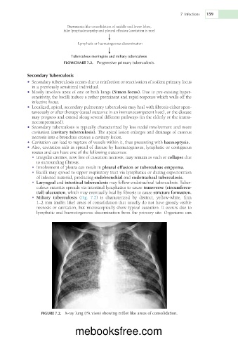
               •  Miliary  tuberculosis  (Fig.  7.2)  is  characterized  by  distinct,  yellow-white,  firm
                 1–2 mm (millet like) areas of consolidation that usually do not have grossly visible
                 necrosis or cavitation, but microscopically show typical caseation. It occurs due to
                 lymphatic and haematogenous dissemination from the primary site. Organisms can


                   FIGURE 7.2.  X-ray lung (PA view) showing millet like areas of consolidation.


                                  mebooksfree.com

