
Page 252 - Concise Pathology for Exam Preparation ( PDFDrive )
P. 252
10 Blood Vessels 237
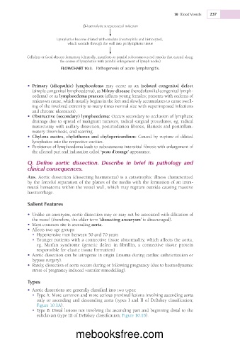
β-haemolytic streptococcal infection
Lymphatics become dilated with exudate (neutrophils and histiocytes),
which extends through the wall into perilymphatic tissue
Cellulitis or focal abscess formation (clinically, manifests as painful subcutaneous red streaks that extend along
the course of lymphatics with painful enlargement of lymph nodes)
FLOWCHART 10.3. Pathogenesis of acute lymphangitis.
• Primary (idiopathic) lymphoedema may occur as an isolated congenital defect
(simple congenital lymphoedema), as Milroy disease (heredofamilial congenital lymph-
oedema) or as lymphoedema praecox (affects young females; presents with oedema of
unknown cause, which usually begins in the feet and slowly accumulates to cause swell-
ing of the involved extremity to many times normal size with superimposed infections
and chronic ulceration).
• Obstructive (secondary) lymphoedema: Occurs secondary to occlusion of lymphatic
drainage due to spread of malignant tumours, radical surgical procedures, eg, radical
mastectomy with axillary dissection, postirradiation fibrosis, filariasis and postinflam-
matory thrombosis, and scarring.
• Chylous ascites, chylothorax and chylopericardium: Caused by rupture of dilated
lymphatics into the respective cavities.
• Persistence of lymphoedema leads to subcutaneous interstitial fibrosis with enlargement of
the affected part and induration called ‘peau d’orange’ appearance.
Q. Define aortic dissection. Describe in brief its pathology and
clinical consequences.
Ans. Aortic dissection (dissecting haematoma) is a catastrophic illness characterized
by the forceful separation of the planes of the media with the formation of an intra-
mural hematoma within the vessel wall, which may rupture outside causing massive
haemorrhage.
Salient Features
• Unlike an aneurysm, aortic dissection may or may not be associated with dilatation of
the vessel (therefore, the older term ‘dissecting aneurysm’ is discouraged).
• Most common site is ascending aorta.
• Affects two age groups:
• Hypertensive men between 50 and 70 years
• Younger patients with a connective tissue abnormality, which affects the aorta,
eg, Marfan syndrome (genetic defect in fibrillin, a connective tissue protein
responsible for elastic tissue formation)
• Aortic dissection can be iatrogenic in origin (trauma during cardiac catheterization or
bypass surgery).
• Rarely, dissection of aorta occurs during or following pregnancy (due to haemodynamic
stress of pregnancy induced vascular remodelling).
Types
• Aortic dissections are generally classified into two types:
• Type A: More common and more serious proximal lesions involving ascending aorta
only or ascending and descending aorta (types I and II of DeBakey classification;
Figure 10.1A).
• Type B: Distal lesions not involving the ascending part and beginning distal to the
subclavian (type III of DeBakey classification; Figure 10.1B).
mebooksfree.com