
Page 254 - Concise Pathology for Exam Preparation ( PDFDrive )
P. 254
10 Blood Vessels 239
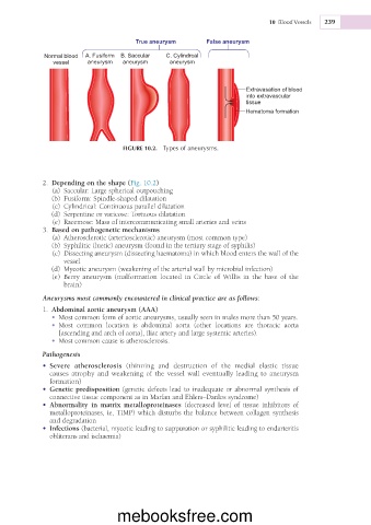
True aneurysm False aneurysm
Normal blood A. Fusiform B. Saccular C. Cylindrcal
vessel aneurysm aneurysm aneurysm
Extravasation of blood
into extravascular
tissue
Hematoma formation
FIGURE 10.2. Types of aneurysms.
2. Depending on the shape (Fig. 10.2)
(a) Saccular: Large spherical outpouching
(b) Fusiform: Spindle-shaped dilatation
(c) Cylindrical: Continuous parallel dilatation
(d) Serpentine or varicose: Tortuous dilatation
(e) Racemose: Mass of intercommunicating small arteries and veins
3. Based on pathogenetic mechanisms
(a) Atherosclerotic (arteriosclerotic) aneurysm (most common type)
(b) Syphilitic (luetic) aneurysm (found in the tertiary stage of syphilis)
(c) Dissecting aneurysm (dissecting haematoma) in which blood enters the wall of the
vessel
(d) Mycotic aneurysm (weakening of the arterial wall by microbial infection)
(e) Berry aneurysm (malformation located in Circle of Willis in the base of the
brain)
Aneurysms most commonly encountered in clinical practice are as follows:
1. Abdominal aortic aneurysm (AAA)
• Most common form of aortic aneurysms, usually seen in males more than 50 years.
• Most common location is abdominal aorta (other locations are thoracic aorta
[ascending and arch of aorta], iliac artery and large systemic arteries).
• Most common cause is atherosclerosis.
Pathogenesis
• Severe atherosclerosis (thinning and destruction of the medial elastic tissue
causes atrophy and weakening of the vessel wall eventually leading to aneurysm
formation)
• Genetic predisposition (genetic defects lead to inadequate or abnormal synthesis of
connective tissue component as in Marfan and Ehlers–Danlos syndrome)
• Abnormality in matrix metalloproteinases (decreased level of tissue inhibitors of
metalloproteinases, ie, TIMP) which disturbs the balance between collagen synthesis
and degradation
• Infections (bacterial, mycotic leading to suppuration or syphilitic leading to endarteritis
obliterans and ischaemia)
mebooksfree.com