
Page 294 - Concise Pathology for Exam Preparation ( PDFDrive )
P. 294
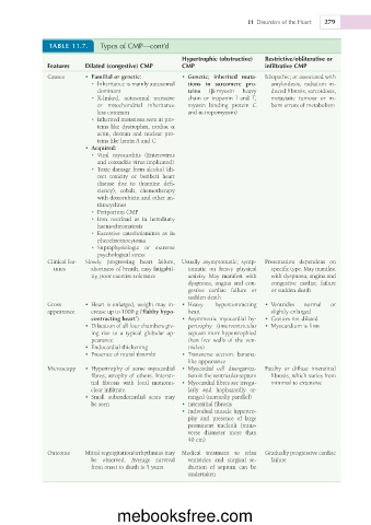
11  Disorders of the Heart  279


               TABLE 11.7.   Types of CMP—cont’d

                                                    Hypertrophic (obstructive)   Restrictive/obliterative or
              Features   Dilated (congestive) CMP   CMP                     infiltrative CMP
              Causes     •  Familial	or	genetic:    •  Genetic;	 inherited	 muta-  Idiopathic; or associated with
                           •  Inheritance is mainly autosomal   tions	 in	 sarcomere	 pro-  amyloidosis,  radiation  in-
                            dominant                  teins   (b-myosin   heavy   duced fibrosis, sarcoidosis,
                           •  X-linked,  autosomal  recessive   chain  or  troponin  I  and  T,   metastatic  tumour  or  in-
                            or  mitochondrial  inheritance   myosin  binding  protein  C   born errors of metabolism
                            less common               and a-tropomyosin)
                           •  Inherited mutations seen in pro-
                            teins like dystrophin, cardiac a
                            actin,  desmin  and  nuclear  pro-
                            teins like lamin A and C
                         •  Acquired:
                           •  Viral  myocarditis  (Enterovirus
                            and coxsackie virus implicated)
                           •  Toxic damage from alcohol (di-
                            rect  toxicity  or  beriberi  heart
                            disease  due  to  thiamine  defi-
                            ciency),  cobalt,  chemotherapy
                            with doxorubicin and other an-
                            thracyclines
                           •  Peripartum CMP
                           •  Iron  overload  as  in  hereditary
                            haemochromatosis
                           •  Excessive  catecholamines  as  in
                            pheochromocytoma
                           •  Supraphysiologic  or  extreme
                            psychological stress
              Clinical fea-  Slowly  progressing  heart  failure,  Usually  asymptomatic;  symp-  Presentation  dependent  on
                tures      shortness of breath, easy fatigabil-  tomatic  on  heavy  physical   specific type. May manifest
                           ity, poor exercise tolerance  activity.  May  manifest  with   with dyspnoea, angina and
                                                      dyspnoea,  angina  and  con-  congestive  cardiac  failure
                                                      gestive  cardiac  failure  or   or sudden death
                                                      sudden death
              Gross      •  Heart is enlarged, weight may in-  •  Heavy   hypercontracting   •  Ventricles   normal   or
              appearance   crease up to 1000 g (‘flabby	hypo-  heart         slightly enlarged
                           contracting	heart’).     •  Asymmetric myocardial hy-  •  Cavities not dilated
                         •  Dilatation of all four chambers giv-  pertrophy  (interventricular   •  Myocardium is firm
                           ing  rise  to  a  typical  globular  ap-  septum more hypertrophied
                           pearance.                  than  free  walls  of  the  ven-
                         •  Endocardial thickening    tricles)
                         •  Presence of mural thrombi   •  Transverse  section:  banana-
                                                      like appearance
              Microscopy  •  Hypertrophy  of  some  myocardial  •  Myocardial  cell  disorganiza-  Patchy  or  diffuse  interstitial
                           fibres; atrophy of others. Intersti-  tion in the ventricular septum  fibrosis, which varies from
                           tial  fibrosis  with  focal  mononu-  •  Myocardial fibres are irregu-  minimal to extensive
                           clear infiltrate.          larly  and  haphazardly  ar-
                         •  Small  subendocardial  scars  may   ranged (normally parallel)
                           be seen                  •  Interstitial fibrosis
                                                    •  Individual muscle hypertro-
                                                      phy  and  presence  of  large
                                                      prominent  nucleoli  (trans-
                                                      verse  diameter  more  than
                                                      40 cm)
              Outcome    Mitral regurgitation/arrhythmias may  Medical  treatment  to  relax   Gradually progressive cardiac
                           be  observed.  Average  survival   ventricles  and  surgical  re-  failure
                           from onset to death is 5 years  duction  of  septum  can  be
                                                      undertaken


                                  mebooksfree.com