
Page 290 - Concise Pathology for Exam Preparation ( PDFDrive )
P. 290
11 Disorders of the Heart 275
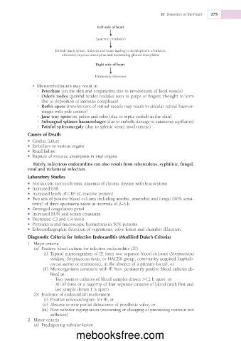
Left side of heart
Systemic circulation
Emboli reach spleen, kidneys and brain leading to development of infarcts,
abscesses, mycotic aneurysms and necrotizing glomerulonephritis
Right side of heart
Pulmonary abscesses
• Microembolization may result in
• Petechiae (on the skin and conjunctiva due to involvement of local vessels)
• Osler’s nodes (painful tender nodules seen in pulps of fingers, thought to form
due to deposition of immune complexes)
• Roth’s spots (involvement of retinal vessels may result in circular retinal haemor-
rhages with pale centres)
• Jane way spots on palms and soles (due to septic emboli in the skin)
• Subungual splinter haemorrhages (due to embolic damage to cutaneous capillaries)
• Painful splenomegaly (due to splenic vessel involvement)
Causes of Death
• Cardiac failure
• Embolism to various organs
• Renal failure
• Rupture of mycotic aneurysms in vital organs
Rarely, infectious endocarditis can also result from tuberculous, syphilitic, fungal,
viral and rickettsial infection.
Laboratory Studies
• Normocytic normochromic anaemia of chronic disease with leucocytosis
• Increased ESR
• Increased levels of CRP (C-reactive protein)
• Two sets of positive blood cultures including aerobic, anaerobic and fungal (90% sensi-
tivity) of three specimens taken at intervals of 2–3 h
• Deranged coagulation panel
• Increased BUN and serum creatinine
• Decreased C3 and C4 levels
• Proteinuria and microscopic haematuria in 50% patients
• Echocardiographic detection of vegetations, valve lesion and chamber dilatation
Diagnostic Criteria for Infective Endocarditis (Modified Duke’s Criteria)
1. Major criteria
(a) Positive blood culture for infective endocarditis (IE)
(i) Typical microorganism of IE from two separate blood cultures (Streptococcus
viridans, Streptococcus bovis, or HACEK group, community acquired Staphylo-
coccus aureus or enterococci, in the absence of a primary focus), or
(ii) Microorganisms consistent with IE from persistently positive blood cultures de-
fined as:
- Two positive cultures of blood samples drawn .12 h apart, or
- All of three or a majority of four separate cultures of blood (with first and
last sample drawn 1 h apart).
(b) Evidence of endocardial involvement
(i) Positive echocardiogram for IE, or
(ii) Abscess or new partial dehiscence of prosthetic valve, or
(iii) New valvular regurgitation (worsening or changing of preexisting murmur not
sufficient)
2. Minor criteria
(a) Predisposing valvular lesion
mebooksfree.com