
Page 348 - Concise Pathology for Exam Preparation ( PDFDrive )
P. 348
12 Haematology 333
Variants of RS Cells
1. Mononuclear variants: Single round to oblong nucleus with a large inclusion-like
nucleolus
2. Lacunar cells: Predominantly seen in NS subtype. Delicate, folded and multilobated
nucleus with abundant pale cytoplasm often disrupted while cutting sections. Nucleus
appears to be sitting in a hole (lacuna).
3. L and H variants:
RS cells undergo mummification (shrinkage and pyknosis) to give rise to cells with
polypoid nuclei resembling popcorn, having inconspicuous nucleoli and moderate
to abundant cytoplasm. Usually seen in the LP subtype.
Note: RS-like cells may be seen in solid cancers, non-Hodgkin lymphoma and infectious
mononucleosis. For diagnosing ‘HL’, RS cells must be present in a background of
non-neoplastic cells (lymphocytes, plasma cells and eosinophils).
Aetiology and Pathogenesis
• The cell of origin of RS cells is thought to be a germinal centre or postgerminal centre
B lymphocyte.
• Rarely (1–2% cases) RS cells have TCR rearrangements suggesting origin from trans-
formed T cells.
• EBV episomes are frequently present in RS cells. EBV-positive tumour cells express
latent membrane protein or LMP-1 (a protein encoded by EBV genome that has
transforming activity).
• LMP-1 upregulates NF-KB (transcription factor responsible for lymphocyte activation).
• NF-KB activation appears to be a common event in classical EBV-positive HL (NF-KB
activation in EBV-negative cases occurs by acquired mutation in a negative regulator
IKB).
• NF-KB activation possibly rescues cells from apoptosis.
• Accumulation of reactive cells is thought to be in response to cytokines released by
RS cells, eg, IL-5, IL-6, IL-13, TNF and GM CSF.
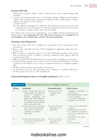
Clinicopathological Features of Hodgkin Lymphoma (Table 12.18)
TABLE 12.18. Clinicopathological features of Hodgkin lymphoma
Subtypes Morphology Immunophenotype Clinical features
NS Frequent ‘lacunar cells’ and occa- RS cells are CD15-and • Mediastinal involvement is
sional diagnostic RS cell; back- 30-positive; EBV- commonly seen
ground of T lymphocytes, eo- negative • Most patients present in Stage
sinophils, macrophages and I or II of the disease
plasma cells. Fibrous bands di- • F 5 M; affects young adults
vide cellular areas into nodules; • Constitutes 65–75% of HL
cells arranged in syncytial sheets
with interspersed necrosis
MC Frequent ‘mononuclear’ and ‘diag- RS cells are CD15-and • . 50% present as Stage III or
nostic RS cells’; background in- 30-positive; 70% IV disease
filtrate rich in T lymphocytes, EBV-positive • Usually involve neck nodes
eosinophils, macrophages, • M . F/biphasic age distribu-
plasma cells tion seen in young adults and
. 55 years
• Constitute 20–25% of HL
LR Frequent ‘mononuclear’ and ‘diag- RS cells are CD15-and • Uncommon
nostic RS cells’, background rich 30-positive; 70% • M . F
in T lymphocytes EBV-positive • Affects older adults
Continued
mebooksfree.com