
Page 350 - Concise Pathology for Exam Preparation ( PDFDrive )
P. 350
12 Haematology 335
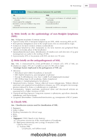
TABLE 12.20. Clinical differences between HL and NHL
HL NHL
More often localized to a single axial group More frequent involvement of multiple periph-
of nodes eral nodes
Orderly spread by contiguity Noncontiguous spread
Mesenteric nodes and Waldeyer ring rarely Commonly involved
involved
Extranodal involvement uncommon Extranodal involvement common
Q. Write briefly on the epidemiology of non-Hodgkin lymphoma
(NHL).
Ans. Malignant neoplasm of immune system:
• Approximately, 60% of malignant lymphomas are NHL, while remaining 40% are HL.
• Most primary malignancies arise in the lymph nodes; few are extranodal in origin.
• Stomach is the most common primary extranodal site.
• Low-grade lymphomas often metastasize to the bone marrow and peripheral blood
(labelled leukaemic phase of the lymphoma).
• Immunohistochemical stains, identification of translocation and detection of Ig gene
rearrangement are useful in the workup of NHL.
• Approximately, 60% of the patients with NHL are men over 50 years.
Q. Write briefly on the aetiopathogenesis of NHL.
Ans. NHL is characterized by clonal proliferation of immune cells. 65% of NHL are
B-lymphocyte origin, 35% are T lymphocyte and 2% NK cell in origin.
Aetiologic factors implicated in the pathogenesis of NHL are
• Infections
• Helicobacter pylori (MALT lymphoma of stomach)
• EBV (Burkitt lymphoma, post-transplant lymphoma)
• Human T-cell leukaemia virus Type I (adult T-cell lymphoma/leukaemia)
• HIV (Diffuse large B-cell lymphoma, Burkitt lymphoma)
• Hepatitis C (Lymphoplasmacytic lymphoma)
• Immunodeficiency diseases: Various inherited (ataxia telangiectasia, Wiskott–Aldrich
syndrome) and acquired immunodeficiency diseases, eg, AIDS, iatrogenic immunosup-
pression induced by chemo or radiotherapy are implicated.
• Autoimmunity: Sjögren syndrome, nontropical sprue and rheumatoid arthritis are
associated with a higher incidence of NHL.
• Chemical and drug exposure: Long-term exposure to phenytoin, agriculture chemicals,
radiotherapy and chemotherapy
• Cytogenetic abnormalities: Chromosomal translocations, eg, overexpression of BCL-2 protein
Q. Classify NHL.
Ans. Classification systems used for classification of NHL:
• Rappaport
• Lukes–Collins
• Working formulation for clinical usage
• REAL
• WHO
1. Rappaport (1966): Based on two features:
(a) Low-power microscopy of the overall pattern of lymphoma
(b) High-power microscopy and cytology of neoplastic cells
Classifies NHLs into:
(i) Nodular NHL
- Lymphocytic, well differentiated
mebooksfree.com