
Page 347 - Concise Pathology for Exam Preparation ( PDFDrive )
P. 347
332 SECTION II Diseases of Organ Systems
Q. Enumerate the causes of generalized lymphadenopathy.
Ans. Causes of generalized lymphadenopathy
• Disseminated tuberculosis
• HIV-associated lymphadenopathy
• Secondary syphilis
• Infectious mononucleosis
• Brucellosis
• Systemic lupus erythaematosus and rheumatoid arthritis
• Lymphomas
• Leukaemias (ALL and CLL)
Neoplastic Proliferations of Lymph Nodes
Q. Write in detail on Hodgkin lymphoma (HL).
Ans. HL has a bimodal age incidence; affects young adults (15–35 years) and older adults
(45–75 years). Reed–Sternberg (RS) cells are the diagnostic hallmark.
Classification
• Nodular sclerosis (NS) • NS, MC, LR & LD are also called “classical HL”.
• Mixed cellularity (MC) • All have RS cells with similar phenotype, positive
• Lymphocyte rich (LR) for PAX5 (a B cell transcription factor), CD15 and
• Lymphocyte depleted (LD) CD30 and negative for other markers.
• Lymphocyte predominant (LP) → B-cell immunophenotype of RS cells (positive for
CD20 and BCL6 and negative for CD15 and CD30).
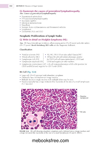
RS Cell (Fig. 12.8)
• Large cell (15–45 microns) with abundant cytoplasm
• Classically has a bilobed mirror image nucleus
• Multiple nuclei or single nucleus with multiple lobes may be seen
• Nucleus typically has a large inclusion-like nucleolus of the size of a small lymphocyte
(5–7 microns)
Reactive
background
RS cells
FIGURE 12.8. RS cell showing abundant cytoplasm and a bilobed mirror image nucleus and
a large inclusion-like nucleolus of the size of a small lymphocyte (H and E; 4003).
mebooksfree.com