
Page 363 - Concise Pathology for Exam Preparation ( PDFDrive )
P. 363
348 SECTION II Diseases of Organ Systems
Laboratory Diagnosis
• Haemogram shows thrombocytopenia with a normocytic normochromic anaemia (con-
sequent to bleeding).
• Peripheral blood smear shows large/giant platelets, reflecting the early release of mega-
karyocytic fragments into the circulation. Platelets lack granules or have an abnormal
colour. Lymphocytosis and eosinophilia are common.
• Tests for antiplatelet antibodies and assays for platelet-associated immunoglobulin, or
antiplatelet antibodies are available.
• Bone marrow shows an increase in the number of megakaryocytes and their precursors
which may show an abnormal morphology. There may be decreased cytoplasmic granu-
larity, variable staining, vacuolization of the cytoplasm and hypolobulation of the nuclei.
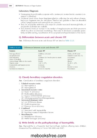
Q. Differentiate between acute and chronic ITP.
Ans. Differences between acute and chronic ITP are listed in Table 12.21.
TABLE 12.21. Differences between acute and chronic ITP
Features Acute ITP Chronic ITP
Peak age affected Children, 2–6 years Adults, 20–40 years
Sex predilection None Female to male ratio 3:1
Prior infection Common (1–3 weeks prior to onset) Uncommon
Onset of bleeding Abrupt Insidious
Haemorrhagic bullae in the mouth Present Absent
9
Platelet count ,20 3 10 /L 30–80 3 10 /L
9
Eosinophilia and lymphocytosis Common Rare
Duration 2–6 weeks Months to years
Spontaneous remission Occurs in majority Uncommon
Q. Classify hereditary coagulation disorders.
Ans. Classification of hereditary coagulation disorders:
1. X-linked recessive traits
(a) Haemophilia A
(b) Haemophilia B
2. Autosomal recessive traits
(a) Factor XI deficiency
(b) Prothrombin deficiency
(c) Factors V/VII/X/XII/XIII deficiency
(d) Afibrinogenaemia/Hypofibrinogenaemia
3. Autosomal dominant traits
(a) von Willebrand disease
(b) Dysfibrinogenaemias
(c) Passovoy factor deficiency
4. Combined
(a) Associated with haemophilia
(b) Involving vitamin K-dependent factors
5. Miscellaneous
(a) Prekallikrein deficiency
(b) HMW Kininogen deficiency
Q. Write briefly on the pathophysiology of haemophilia.
Ans. Haemophilia is a frequently fatal haemorrhagic diathesis affecting male children
characterized by a deficiency of Factor VIII (AHG and AHF).
mebooksfree.com