
Page 403 - Concise Pathology for Exam Preparation ( PDFDrive )
P. 403
388
388 SECTION II Diseases of Organ Systems
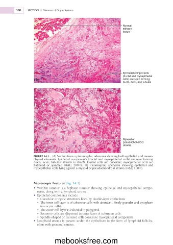
Normal
salivary
tissue
Epithelial components
(ductal and myoepithelial
A cells) are seen forming
ducts, acini, and tubules
Myxoid or
pseudochondroid
stroma
B
FIGURE 14.1. (A) Section from a pleomorphic adenoma showing both epithelial and mesen-
chymal elements. Epithelial components (ductal and myoepithelial cells) are seen forming
ducts, acini, tubules, strands or sheets. Ductal cells are cuboidal; myoepithelial cells are
flattened or spindled (H&E; 2003). (B) Pleomorphic adenoma showing epithelial and
myoepithelial cells lying against a myxoid or pseudochondroid stroma (H&E; 1003).
Microscopic Features (Fig. 14.2)
• Warthin tumour is a biphasic tumour showing epithelial and myoepithelial compo-
nents, along with a lymphoid stroma.
• Epithelial components include
• Glandular or cystic structures lined by double-layer epithelium
• The inner cell layer is of columnar cells with abundant, finely granular and cytoplasm
(oncocytic cells).
• The outer cell layer is cuboidal to polygonal.
• Secretory cells are dispersed in inner layer of columnar cells.
• Spindle-shaped or flattened cells constitute myoepithelial component
• Lymphoid stroma is present under the epithelium in the form of lymphoid follicles,
often with germinal centres.
mebooksfree.com