
Page 404 - Concise Pathology for Exam Preparation ( PDFDrive )
P. 404
14 The Oral Cavity and Gastrointestinal Tract 389
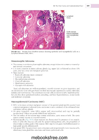
Lymphoid
tissue
Cystic spaces
lined by epithelial
and myoepithelial
cells
FIGURE 14.2. Section from Warthin tumour showing epithelial and myoepithelial cells in a
lymphoid stroma (H&E; 403).
Monomorphic Adenoma
• This tumour is similar to pleomorphic adenoma, except it does not contain a mesenchy-
mal stromal component.
• It is more common in minor salivary glands (eg, upper lip), is bilateral in about 10%
cases, and has a very rare malignant potential.
• Types include
• Basal-cell adenoma (most common)
• Canicular adenoma
• Myoepithelioma adenoma
• Clear-cell adenoma
• Membranous adenoma
• Glycogen-rich adenoma
Basal cell adenomas are well-encapsulated, smooth tumours on gross inspection, and
are divided into four subtypes based on their microscopic appearance—solid, trabecular,
tubular and membranous. The constituent tumour cells are monomorphic basaloid epithe-
lial cells that show peripheral nuclear palisading, and have hyperchromatic, round nuclei
and indistinct cytoplasm.
Mucoepidermoid Carcinoma (MEC)
• MEC is the most common malignant tumour of the parotid gland and the second most
common malignancy (adenoid cystic carcinoma is more common) of the submandibular
and minor salivary glands.
• On gross inspection, some MECs appear well circumscribed and may be partially
encapsulated. Others are poorly defined and infiltrative.
• The cut surface of the tumour may contain solid areas, cystic areas or both. The cystic
spaces contain viscous or mucoid material.
• Microscopically (Fig. 14.3); these tumours are characterized by presence of two popula-
tions of cells—the mucous cells and the epidermoid/squamous cells, the proportion of
which helps to define grade of the tumour. MEC may be low grade (well differentiated) or
high grade (poorly differentiated).
• Low-grade MEC has prominent cystic structures and proportionally more mucous cells,
which may form gland-like structures and fewer epidermoid cells.
mebooksfree.com