
Page 405 - Concise Pathology for Exam Preparation ( PDFDrive )
P. 405
390
390 SECTION II Diseases of Organ Systems
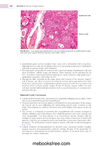
Epidermoid cells
Mucous cells
FIGURE 14.3. Low-grade mucoepidermoid carcinoma displaying both an epidermoid compo-
nent and cystic spaces lined by mucous cells (H&E; 2003).
• Intermediate-grade tumours display fewer cysts and a substantial solid component.
Although mucous cells are still present, there is an increasing proportion of epidermoid
cells and occasional keratin pearl formation.
• The high-grade carcinomas are solid tumours comprised mainly of epidermoid cells that
show prominent cellular atypia and mitoses. These tumours can be mistaken for an
SCC. A positive immunohistochemical staining for mucin indicates a high-grade muco-
epidermoid carcinoma, rather than an SCC.
• Therapy for MEC depends on the stage, grade and location of the tumour. Stages I
and II disease can often be treated by surgical excision alone (parotidectomy with
facial nerve preservation, submandibular gland excision or wide local excision of an
involved minor salivary gland). Stages III and IV disease require radical excision
and may warrant additional intervention such as a neck dissection or postoperative
radiation therapy.
Adenoid Cystic Carcinoma
• It peaks in fifth decade of life, and presents as a gradually enlarging salivary mass, which
may be accompanied by pain and paraesthesias.
• On gross inspection the tumour appears well defined but unencapsulated. In late stages,
the tumour can be seen infiltrating the surrounding normal tissue. Contrary to the
name, these tumours are solid in consistency and rarely display cystic spaces on the cut
surface.
• The tumour is composed of epithelial and myoepithelial cells variably arranged in
tubular, cribriform and solid patterns. The cribriform pattern is the most common and
easily recognizable. It is often referred to as ‘Swiss-cheese’ pattern. Tumour cells are
arranged in nests around cylindrical spaces that may contain a mucinous or hyalinized
material. Cells that are arranged in layers and form ductal structures characterize tubu-
lar pattern. The solid pattern contains sheets of tumour cells with no intervening spaces
(Fig. 14.4).
• Current treatment recommendations for adenoid cystic carcinoma include complete
surgical resection and postoperative radiation therapy. Because of the propensity for
this tumour to demonstrate perineural invasion, sacrifice of the facial nerve may be
necessary for tumour eradication.
mebooksfree.com