
Page 470 - Concise Pathology for Exam Preparation ( PDFDrive )
P. 470
16 Diseases of the Kidney and Lower Urinary Tract 455
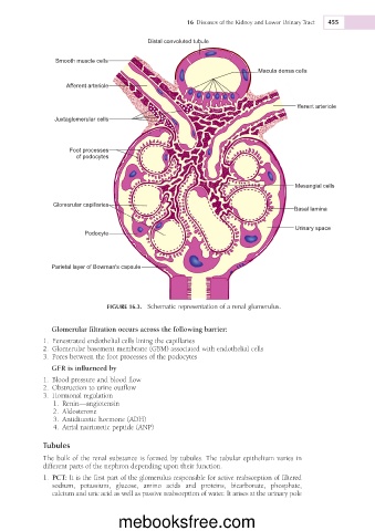
Distal convoluted tubule
Smooth muscle cells
Macula densa cells
Afferent arteriole
Efferent arteriole
Juxtaglomerular cells
Foot processes
of podocytes
Mesangial cells
Glomerular capillaries
Basal lamina
Urinary space
Podocyte
Parietal layer of Bowman's capsule
FIGURE 16.3. Schematic representation of a renal glomerulus.
Glomerular filtration occurs across the following barrier:
1. Fenestrated endothelial cells lining the capillaries
2. Glomerular basement membrane (GBM) associated with endothelial cells
3. Pores between the foot processes of the podocytes
GFR is influenced by
1. Blood pressure and blood flow
2. Obstruction to urine outflow
3. Hormonal regulation
1. Renin—angiotensin
2. Aldosterone
3. Antidiuretic hormone (ADH)
4. Atrial natriuretic peptide (ANP)
Tubules
The bulk of the renal substance is formed by tubules. The tubular epithelium varies in
different parts of the nephron depending upon their function.
1. PCT: It is the first part of the glomerulus responsible for active reabsorption of filtered
sodium, potassium, glucose, amino acids and proteins, bicarbonate, phosphate,
calcium and uric acid as well as passive reabsorption of water. It arises at the urinary pole
mebooksfree.com