
Page 468 - Concise Pathology for Exam Preparation ( PDFDrive )
P. 468
16
Diseases of the Kidney and
Lower Urinary Tract
NORMAL STRUCTURE
The kidneys are paired, bean-shaped, retroperitoneal organs each weighing about 150 g
in the adult male and 135 g in the adult female. They are typically 10–12 cm in length,
5–7 cm in width and 2–3 cm in thickness. The renal artery, vein, lymphatics and
the ureters are located in the renal hilum which is the centre of the concave area of the
kidney. The upper and lower poles of each kidney lie opposite to the twelfth thoracic
vertebra, and the third lumbar vertebra, respectively. Right kidney is slightly lower due
to the presence of liver. The renal capsule is a smooth, transparent, fibrous membrane
that is normally easily removable. It protects the organ and is surrounded by peri-
renal fat which further cushions the kidneys.
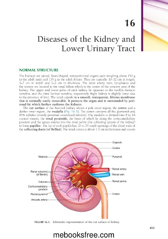
The cut surface of the bisected kidney shows a pale outer region, the cortex and a
darker inner region, the medulla (Fig. 16.1). The cortex contains all the glomeruli and
85% tubules (mainly proximal convoluted tubules). The medulla is divided into 8 to 18
conical masses, the renal pyramids, the bases of which lie along the corticomedullary
junction and the apices extend into the renal pelvis (the collecting system of the kidney)
to form papillae. The tip of each papilla has 10 to 25 small openings of the distal ends of
the collecting ducts (of Bellini). The renal cortex is about 1.5 cm in thickness and covers
FIGURE 16.1. Schematic representation of the cut surface of kidney.
453
mebooksfree.com