
Page 469 - Concise Pathology for Exam Preparation ( PDFDrive )
P. 469
454 SECTION II Diseases of Organ Systems
Glomerulus Distal convoluted
tubule
Proximal
convoluted Bowman’s Cortical
tubule capsule collecting
duct
Thick
ascending
Cortex limb
Collecting
Outer medulla duct
Thin Thin
descending ascending
limb
Inner limb
medulla Inner medulla
(papilla) collecting duct
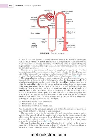
FIGURE 16.2. Parts of a nephron.
the base of each renal pyramid to extend downward between the individual pyramids to
form the renal columns of Bertin. The ureter on entering the kidney dilates to form the
renal pelvis which is lined by transitional epithelium and forms 2–3 outpouchings, the
major calyces. From each of the major calyces, several minor calyces extend toward the
papillae of the pyramids.
The main unit of parenchyma of each kidney is the nephron. There are about 1–4 million
nephrons in each kidney. Each nephron contains 5 major subunits, the dilated ‘glomerulus
with the Bowman capsule’, ‘the proximal convoluted tubule or PCT’, ‘the thin and thick loop
of Henle’, ‘the distal convoluted tubule’ or DCT and the ‘collecting ducts’ (Fig. 16.2).
The glomerulus is a bulbous structure invaginated by a capillary network which is
surrounded by a double-layered epithelial capsule called the Bowman’s capsule. The
inner layer enveloping the capillary tuft is called the visceral layer and the outer layer is
called the parietal layer. Between the visceral and the parietal epithelial layers is a cavity
called Bowman’s space. The large area of the capillary network makes the glomerulus
an efficient filtration unit. Each nephron has a vascular pole and a urinary pole. The
vascular pole is where the afferent arteriole enters and the efferent arteriole leaves.
The PCT begins at the urinary pole. The inner side of the glomerular capillary wall
is lined by a thin layer of fenestrated endothelial cells which rest on the glomerular
basement membrane (GBM). The GBM is constituted by collagen, laminin, fibronectin,
proteoglycans and glycoproteins and has three layers:
(a) Lamina rara externa on the external side
(b) Lamina densa in the middle
(c) Lamina rara interna on the internal side
Any abnormality in the glomerular epithelial cells or the above-mentioned three layers
may disturb the barrier to filtration of macromolecules.
The glomerulus is supported by mesangial cells with surrounding mesangial matrix
material. The external side of the capillary wall is lined by the visceral epithelial cells
which rest on the lamina rara externa. These cells have foot-like extensions and are there-
fore also called podocytes (podo-foot). The podocytes have 20–30 nm wide spaces
between them to allow filtration (Fig. 16.3).
The main function of the glomerulus is filtration from the capillaries to the Bowman’s
space. Normal glomerular filtration rate is about 125 mL/min. The glomerular filtrate is
identical to plasma in composition except it lacks cells and protein.
mebooksfree.com