
Page 551 - Concise Pathology for Exam Preparation ( PDFDrive )
P. 551
536 SECTION II Diseases of Organ Systems
supported by the fact that there is a 60% concordance in monozygotic twins and an as-
sociation with HLA B8 and DR3. The genetic susceptibility is linked to polymorphisms
in multiple immune regulatory genes, eg, cytotoxic T-lymphocyte-associated antigen
4 (CTLA 4) and protein tyrosine phosphatase 22 (PTPN 22). Graves disease is a triad of:
• Hyperthyroidism due to diffuse hyperplasia of follicular epithelium
• Infiltrative ophthalmopathy with resultant exophthalmos
• Localized infiltrative dermopathy called pretibial myxoedema
Pathogenesis
Multiple autoantibodies have been demonstrated in Graves disease, primarily
against the TSH receptor. These include:
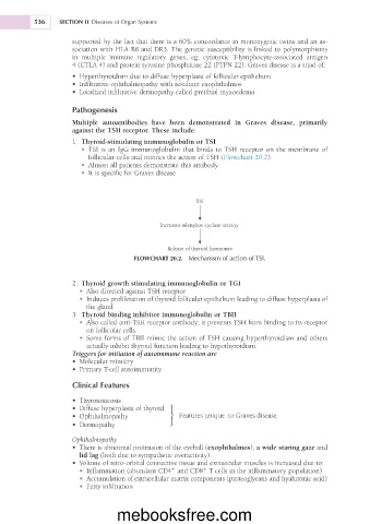
1. Thyroid-stimulating immunoglobulin or TSI
• TSI is an IgG immunoglobulin that binds to TSH receptor on the membrane of
follicular cells and mimics the action of TSH (Flowchart 20.2)
• Almost all patients demonstrate this antibody
• It is specific for Graves disease
TSI
Increases adenylate cyclase activity
Release of thyroid hormones
FLOWCHART 20.2. Mechanism of action of TSI.
2. Thyroid growth stimulating immunoglobulin or TGI
• Also directed against TSH receptor
• Induces proliferation of thyroid follicular epithelium leading to diffuse hyperplasia of
the gland
3. Thyroid binding inhibitor immunoglobulin or TBII
• Also called anti-TSH receptor antibody; it prevents TSH from binding to its receptor
on follicular cells.
• Some forms of TBII mimic the action of TSH causing hyperthyroidism and others
actually inhibit thyroid function leading to hypothyroidism.
Triggers for initiation of autoimmune reaction are
• Molecular mimicry
• Primary T-cell autoimmunity
Clinical Features
• Thyrotoxicosis
• Diffuse hyperplasia of thyroid
• Ophthalmopathy } Features unique to Graves disease
• Dermopathy
Ophthalmopathy
• There is abnormal protrusion of the eyeball (exophthalmos), a wide staring gaze and
lid lag (both due to sympathetic overactivity).
• Volume of retro-orbital connective tissue and extraocular muscles is increased due to:
1
1
• Inflammation (abundant CD4 and CD8 T cells in the inflammatory population)
• Accumulation of extracellular matrix components (proteoglycans and hyaluronic acid)
• Fatty infiltration
mebooksfree.com