
Page 555 - Concise Pathology for Exam Preparation ( PDFDrive )
P. 555
540 SECTION II Diseases of Organ Systems
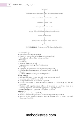
Viral infection
Provision of antigen (viral antigen or virus-induced/altered host antigen)
Antigen presentation in association with HLA-B35
Formation of cytotoxic T cells
Damage to thyroid follicular cells
Rupture of thyroid follicles and release of thyroid hormones
Transient hyperthyroidism
Hypothyroidism (due to loss of thyroid substance)
Recovery
FLOWCHART 20.4. Pathogenesis of de Quervain thyroiditis.
Gross morphology:
• Unilateral or bilateral enlargement
• Capsule is intact and may be adherent to surroundings
• Cut surface is yellow-white, rubbery and firm
Microscopy:
Early changes:
• Scattered disruption of follicles
• Replacement by neutrophilic microabscesses
Late changes:
• Aggregates of lymphocytes, histiocytes and plasma cells
• Presence of multinucleate giant cells around pools of colloid
• Fibrosis
(c) Subacute lymphocytic (painless) thyroiditis
Salient features:
• Affects middle-aged women generally in the postpartum period
• Associated with HLA-DR3 and -DR5
• Thought to be variant of Hashimoto thyroiditis
• Patients demonstrate increased levels of antibodies to thyroglobulin and thyroid
peroxidase
• Manifests with hyperthyroidism followed by reversion to a euthyroid state. In a
minority of patients, the disease may progress to hypothyroidism.
Gross morphology: Mild symmetric enlargement of thyroid
Microscopy:
• Focal disruption of thyroid follicles
• Multifocal inflammatory infiltrate (predominantly small lymphocytes)
• No plasma cells, germinal centres or Hürthle cell metaplasia. If present, think of
Hashimoto thyroiditis.
mebooksfree.com