
Page 611 - Concise Pathology for Exam Preparation ( PDFDrive )
P. 611
596 SECTION II Diseases of Organ Systems
• Peripheral neuropathy
• Pulmonary involvement (Pleuritis, intrapulmonary nodules, interstitial fibrosis in the
form of Caplan syndrome—restrictive lung disease with rheumatoid nodules and coal
worker’s pneumoconiosis)
• Hepatitis
• Ocular involvement (scleritis, episcleritis and dryness of the eye)
• Secondary amyloidosis and Sjögren syndrome
Pathogenesis
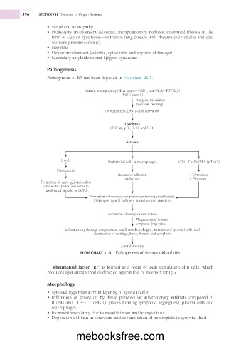
Pathogenesis of RA has been depicted in Flowchart 21.5.
Genetic susceptibility (HLA genes---DRB1; non-HLA---PTPN22)
(MHC class II)
Antigenic stimulation
(infection, smoking)
Unregulated CD4+ T-cells activation
Cytokines
(TNF-α, γ-IF, IL-17 and IL-1)
Activate
B cells
Endothelial cells & macrophages CD4+ T cells (Th1 & Th17)
Plasma cells
Release of adhesion • Cytokines
molecules • Proteases
Formation of Anti-IgG antibodies
(rheumatoid factor, antibodies to
citrullinated peptides or CCPs)
Formation of immune complexes containing citrullinated
fibrinogen, type II collagen, α-enolase and vimentin
Activation of complement system
Phagocytosis of immune
complexes (ragocytes)
Inflammatory damage to synovium, small vessels, collagen, activation of synovial cells, and
destruction of cartilage, bone, fibrosis and ankylosis
Joint deformity
FLOWCHART 21.5. Pathogenesis of rheumatoid arthritis
Rheumatoid factor (RF) is formed as a result of local stimulation of B cells, which
produces IgM autoantibodies directed against the Fc receptor for IgG.
Morphology
• Synovial hyperplasia (multilayering of synovial cells)
• Infiltration of synovium by dense perivascular inflammatory infiltrate composed of
B cells and CD41 T cells (at places forming lymphoid aggregates), plasma cells and
macrophages
• Increased vascularity due to vasodilatation and telangiectasia
• Deposition of fibrin in synovium and accumulation of neutrophils in synovial fluid
mebooksfree.com