
Page 625 - Concise Pathology for Exam Preparation ( PDFDrive )
P. 625
610 SECTION II Diseases of Organ Systems
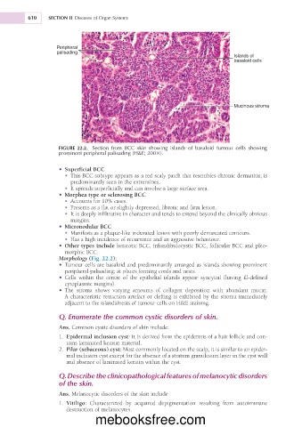
Peripheral
palisading
Islands of
basaloid cells
Mucinous stroma
FIGURE 22.2. Section from BCC skin showing islands of basaloid tumour cells showing
prominent peripheral palisading (H&E; 2003).
• Superficial BCC
• This BCC subtype appears as a red scaly patch that resembles chronic dermatitis; is
predominantly seen in the extremities.
• It spreads superficially and can involve a large surface area.
• Morphea type or sclerosing BCC
• Accounts for 10% cases.
• Presents as a flat or slightly depressed, fibrotic and firm lesion.
• It is deeply infiltrative in character and tends to extend beyond the clinically obvious
margins.
• Micronodular BCC
• Manifests as a plaque-like indurated lesion with poorly demarcated contours.
• Has a high incidence of recurrence and an aggressive behaviour.
• Other types include keratotic BCC, infundibulocystic BCC, follicular BCC and pleo-
morphic BCC.
Morphology (Fig. 22.2):
• Tumour cells are basaloid and predominantly arranged as islands showing prominent
peripheral palisading; at places forming cords and nests.
• Cells within the centre of the epithelial islands appear syncytial (having ill-defined
cytoplasmic margins).
• The stroma shows varying amounts of collagen deposition with abundant mucin.
A characteristic retraction artefact or clefting is exhibited by the stroma immediately
adjacent to the islands/nests of tumour cells on H&E staining.
Q. Enumerate the common cystic disorders of skin.
Ans. Common cystic disorders of skin include:
1. Epidermal inclusion cyst: It is derived from the epidermis of a hair follicle and con-
tains laminated keratin material.
2. Pilar (sebaceous) cyst: Most commonly located on the scalp, it is similar to an epider-
mal inclusion cyst except for the absence of a stratum granulosum layer in the cyst wall
and absence of laminated keratin within the cyst.
Q. Describe the clinicopathological features of melanocytic disorders
of the skin.
Ans. Melanocytic disorders of the skin include
1. Vitiligo: Characterized by acquired depigmentation resulting from autoimmune
destruction of melanocytes.
mebooksfree.com