
Page 627 - Concise Pathology for Exam Preparation ( PDFDrive )
P. 627
612 SECTION II Diseases of Organ Systems
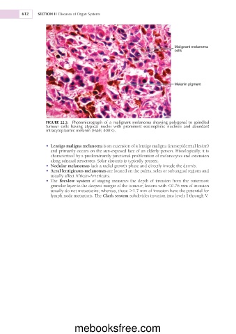
Malignant melanoma
cells
Melanin pigment
FIGURE 22.3. Photomicrograph of a malignant melanoma showing polygonal to spindled
tumour cells having atypical nuclei with prominent eosinophilic nucleoli and abundant
intracytoplasmic melanin (H&E; 4003).
• Lentigo maligna melanoma is an extension of a lentigo maligna (intraepidermal lesion)
and primarily occurs on the sun-exposed face of an elderly person. Histologically, it is
characterized by a predominantly junctional proliferation of melanocytes and extension
along adnexal structures. Solar elastosis is typically present.
• Nodular melanomas lack a radial growth phase and directly invade the dermis.
• Acral lentiginous melanomas are located on the palms, soles or subungual regions and
usually affect African-Americans.
• The Breslow system of staging measures the depth of invasion from the outermost
granular layer to the deepest margin of the tumour; lesions with ,0.76 mm of invasion
usually do not metastasize; whereas, those .1.7 mm of invasion have the potential for
lymph node metastasis. The Clark system subdivides invasion into levels I through V.
mebooksfree.com