
Page 629 - Concise Pathology for Exam Preparation ( PDFDrive )
P. 629
614 SECTION II Diseases of Organ Systems
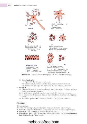
FIGURE 23.1. Normal cells constituting CNS and their cellular morphology.
Ependymal cells
(c)
(i) ‘Epithelial-like’ cuboidal to columnar
(ii) Ciliated luminal surface (cilia for circulation of cerebrospinal fluid)
(iii) Bases of the cells taper and breakdown into fine ramifying processes
Microglia
2.
(a)
Small mobile cells of mesenchymal origin found throughout the brain, predomi-
nantly in perivascular location.
(b)
Irregular nuclei with little cytoplasm and tiny, highly branched processes.
(c)
In response to inflammation, they are transformed into amoeboid cells for phago-
cytosis.
(d) cells’ (due to the presence of phagocytosed material).
Also called ‘glitter
Meninges
Leptomeninges:
The inner two meninges, arachnoid and pia mater, constitute the leptomeninges.
• Piamater: Composed of fibroblasts, collagen and processes of underlying astrocytes.
• Arachnoid: Lined by a layer of flattened mesothelial cells.
• Subarachnoid space: Space between the two leptomeninges; contains cerebrospinal
fluid (CSF) and major blood vessels.
mebooksfree.com