
Page 1210 - Hematology_ Basic Principles and Practice ( PDFDrive )
P. 1210
1056 Part VII Hematologic Malignancies
transduction–transplantation CML model has been helpful in study-
ing the role of BCR-ABL domains and signaling interaction in primary
hematopoietic cells in vivo. The ABL tyrosine kinase is crucial for
oncogenic transformation. Mice that express a form of BCR-ABL
with a point mutation in the ATP-binding site of ABL that inhibits
its kinase activity do not develop leukemia, suggesting that the ABL
kinase activity is essential for BCR-ABL leukemogenesis in vivo. The
success of kinase inhibitor therapy for CML provides further proof of
the importance of kinase activity in maintenance of human disease.
Other important domains in BCR-ABL also regulate the kinase
9 22
activity of ABL or connect to other downstream signaling pathways.
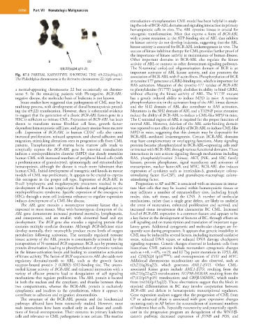
t(9;22)(q34;q11.2) The N-terminal coiled-coil oligomerization domain of BCR is an
important activator of ABL kinase activity, and also promotes the
Fig. 67.1 PARTIAL KARYOTYPE SHOWING THE t(9;22)(q34;q11). association of BCR-ABL with F-actin fibers. Phosphorylation of BCR
The Philadelphia chromosome is the derivative chromosome 22 (right arrow).
at tyrosine 177 generates a GRB2-binding site, which is important for
RAS activation. Mutation of the tyrosine-177 residue of BCR-ABL
a normal-appearing chromosome 22 but occasionally on chromo- to phenylalanine (Y177F) largely abolishes its ability to bind GRB2,
some 9. In the remaining patients with Ph-negative, BCR-ABL- without affecting the kinase activity of ABL. The Y177F mutant
negative disease, the molecular basis of leukemia is not known. has a greatly reduced ability to induce MPD in mice. A tyrosine
Some studies have suggested that pathogenesis of CML may be a phosphorylation site in the activation loop of the ABL kinase domain
multistep process, with development of clonal hematopoiesis preced- and the SH2 domain of ABL also contribute to RAS activation.
ing the t(9;22) translocation. However, there is substantial evidence Mutations in the SH2 domain of ABL and a Y1294F point mutation
to suggest that the generation of a classic BCR-ABL fusion gene in a reduce the ability of BCR-ABL to induce a CML-like MPD in mice.
HSC is sufficient to initiate CML. Expression of BCR-ABL has been The C-terminal region of ABL is required for the proper function of
shown to transform mouse fibroblast cell lines, growth factor- normal ABL. However, deletion of the ABL actin-binding domain
dependent hematopoietic cell lines, and primary murine bone marrow was reported to not affect the ability of BCR-ABL to induce CML-like
+
cells. Expression of BCR-ABL in human CD34 cells also causes MPD in mice, suggesting that this domain may be dispensable for
increased proliferation, reduced apoptosis, and altered adhesion and BCR-ABL–mediated leukemogenesis. Certain BCR-ABL domains
migration, mimicking alterations seen in progenitor cells from CML may have complementary or overlapping functions. Many signaling
patients. Transplantation of murine bone marrow cells made to proteins become phosphorylated in BCR-ABL–expressing cells and/
ectopically express the BCR-ABL gene by retroviral transduction or interact with BCR-ABL through various functional domains. These
induces a myeloproliferative disorder (MPD) that closely resembles interactions in turn activate signaling through mechanisms including
human CML with increased numbers of peripheral blood cells (with RAS, phosphatidylinositol 3-kinase, AKT, JNK, and SRC family
a predominance of granulocytes), splenomegaly, and extramedullary kinases, protein phosphatase, signal transducers and activators of
hematopoiesis, although the disease is much more fulminant than transcription, nuclear factor-κB, and MYC. BCR-ABL also induces
human CML. Initial development of transgenic and knock-in mouse expression of cytokines such as interleukin-3, granulocyte colony-
models of CML was problematic. It appears to be crucial to express stimulating factor (G-CSF), and granulocyte-macrophage colony-
this oncogene in the proper cell type. Expression of BCR-ABL in stimulating factor.
B-cell lymphocytic and megakaryocytic precursors resulted in the Progression to AP and BC is associated with an increase in imma-
development of B-acute lymphocytic leukemia and megakaryocytic ture blast cells that may be located within hematopoietic tissues or
myeloproliferative syndrome. Specific expression of the oncogene in may infiltrate a number of extramedullary sites, including lymph
HSCs through a stem cell leukemia enhancer to regulate expression nodes, skin, soft tissue, and the CNS. A number of molecular
induces development of a CML-like disease. mechanisms, rather than a single gene defect, are likely to underlie
The ABL gene encodes a nonreceptor tyrosine kinase that is the arrest of maturation, enhanced proliferation and survival, and
expressed in most tissues. Mice with homozygous disruption of the increased tissue invasiveness that characterize BC CML. Increased
ABL gene demonstrate increased perinatal mortality, lymphopenia, level of BCR-ABL expression is a common feature and appears to be
and osteoporosis, and are smaller, with abnormal head and eye a key factor in the development of features of BC, through effects on
development. The BCR gene also encodes a signaling protein that cell signaling and on transcription and translation of important regu-
contains multiple modular domains. Although BCR-deficient mice latory genes. Additional cytogenetic and molecular changes are fre-
develop normally, their neutrophils produce excess levels of oxygen quently seen during progression. It appears that genetic instability in
metabolites following activation. The normally regulated tyrosine CML may be induced by several factors, including increased oxidative
kinase activity of the ABL protein is constitutively activated by the stress, reduced DNA repair, or reduced DNA damage checkpoint
juxtaposition of N-terminal BCR sequences. BCR acts by promoting signaling response. Genetic changes observed in leukemic cells from
protein dimerization, leading to phosphorylation of tyrosine residues blast-phase CML patients include nonrandom cytogenetic changes
in the kinase-activation loops and leading to constitutive activation such as ++8, ++Ph, ++19, and I(17)q; point mutations in TP53, RB,
of kinase activity. The fusion of BCR sequences to ABL also adds new and CDKN2A (p16 INK4A ); and overexpression of EVI1 and MYC.
regulatory domains/motifs to ABL, such as the growth factor Additional chromosome translocations are also observed, such as
receptor-bound protein 2 (GRB2) SH2-binding site. The uncon- t(3;21)(q26;q22), which generates AML1-EVI1. Other CML-
trolled kinase activity of BCR-ABL and enhanced interaction with a associated fusion genes include AML1-ETO, resulting from the
variety of effector proteins lead to deregulation of cell signaling t(8;21)(q22;q22) translocation; NUP98-HOXA9, resulting from the
mechanisms that regulate proliferation. The ABL protein is located t(7;11)(p15;p15) translocation; and CBFβ-SMMHC, which results
in both the nucleus and the cytoplasm, and shuttles between these from inv(16)(p13;q22). These observations suggest that the block in
two compartments, whereas the BCR-ABL protein is exclusively myeloid differentiation in BC may involve cooperation between
cytoplasmic and localizes to the cytoskeleton, where it appears to BCR-ABL and defects in hematopoietic transcriptional regulators.
contribute to adhesion and migration abnormalities. Gene expression analyses suggest that the progression of CML from
The structure of the BCR-ABL protein and the biochemical CP to advanced phase is associated with gene expression changes
pathways affected have been extensively studied. However, most occurring early in AP before the accumulation of increased numbers
such interactions have been studied only in cell lines and condi- of leukemia blast cells. Especially noteworthy and potentially signifi-
tions of forced overexpression. Their existence in primary leukemia cant in the progression program are deregulation of the WNT/β-
cells and relevance to CML pathogenesis is not certain. The murine catenin pathway, decreased expression of JUNB and FOS, and