
Page 1228 - Hematology_ Basic Principles and Practice ( PDFDrive )
P. 1228
1074 Part VII Hematologic Malignancies
received a renal allograft. The pathogenesis of this anemia is not clear, somatic or inherited germ-line mutations expressed by hematopoietic
but reduced levels of circulating EPO are not solely responsible, progenitor cells (HPCs). In contrast, secondary polycythemias are
suggesting that there might be other contributing factors. The AT1 characterized by an increase in regulatory growth factors, primarily
receptor is present on erythroid progenitors, and its ligand, AngII, EPO, and normal responsiveness of their erythroid progenitors to
augments EPO stimulation of erythropoiesis. The involvement of these growth factors. These conditions can usually be distinguished
JAK2 kinase in AngII signaling suggests that this signal transduction by in vitro assays of erythroid progenitor cells, quantitation of serum
pathway mediated by EPO and AngII might overlap. Postrenal EPO levels, and detection of somatic JAK2 mutations. In a small
transplant erythrocytosis likely can be accounted for by activation of number of patients, the cause of erythrocytosis cannot be determined;
the RAS. these patients are classified as having idiopathic erythrocytosis.
DEFINITION AND CLASSIFICATION OF POLYCYTHEMIA RELATIVE POLYCYTHEMIA
The term polycythemia is a literal translation from Greek, meaning Individuals with a modestly increased venous hematocrit level that is
“too many cells in the blood,” and refers to an increase in the RBC not accompanied by an increased RBC mass are frequently thought
mass; it is frequently used interchangeably with the term erythrocytosis. to be polycythemic by imprecise yet widely accepted medical practice.
Polycythemia may be due to a myriad of causes (Table 68.1). The Frequently, these individuals are thought to be polycythemic owing
polycythemias can be classified as relative and absolute. Relative to the lack of appreciation by a clinician of what constitutes the upper
polycythemia is a disorder in which the patient characteristically has limit of normal values for a hematocrit (49% in males and 48% in
a modest elevation of the hematocrit level without an elevated RBC females). Such individuals frequently prove not to have an absolute
mass but rather because of contraction of the plasma volume. The polycythemia as defined by an actual increase in the measured RBC
absolute polycythemias are accompanied by an actual increase in the mass. Relative or spurious polycythemia is a term used to describe an
circulating RBC mass. Polycythemias can also be classified according elevation of the hematocrit level either caused by an acute transient
to the responsiveness of their erythroid progenitor cells to growth state of hemoconcentration associated with intravascular fluid deple-
factors or the circulating levels of such growth factors. Primary tion or a chronic sustained relative polycythemia caused by contrac-
polycythemias are characterized by increased sensitivity of the ery- tion of the plasma volume (see Table 68.1).
throid progenitors to regulatory growth factors as a result of acquired Transient polycythemias may be a result of acute depletion of the
plasma volume from a variety of disorders, including protracted
vomiting or diarrhea, plasma loss from external burns, sudden cold
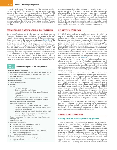
TABLE Differential Diagnosis of the Polycythemias exposure or protracted exercise, insensible fluid loss from fever, sepsis,
68.1 diabetic ketoacidosis, or acute ethanol intoxication. These elevations
of hematocrit can be easily corrected by appropriate replacement of
Relative or Spurious Polycythemia intravascular fluids.
1. Decreased plasma volume—reduced fluid intake, marked loss of Gaisböck syndrome, first described in 1905, is a condition
body fluids (diaphoresis, vomiting, diarrhea, “third spacing”) observed mainly in obese, hypertensive, middle-aged, male smokers.
2. Gaisböck syndrome Alcohol, diuretics, obesity, hypoxia, psychologic stress, and excess
3. Overfilling of blood in collection vacuum tubes catecholamine secretion have been identified as possible causes of
Absolute Polycythemia relative polycythemia. Such individuals can have a chronic modest-
1. Secondary polycythemia to-moderate elevation of the hematocrit level associated with a
A. Acquired normal RBC mass and low plasma volume, which has been attributed
Hypoxia to reduced venous compliance, or they can have a high normal RBC
• Pulmonary disease mass with either a normal or slightly decreased plasma volume. The
• Cyanotic congenital heart disease primary significance of the identification of a patient with relative
• Hypoventilation syndromes: sleep apnea, Pickwickian polycythemia is the recognition of the increased risk of developing
syndrome thrombotic vascular events likely caused by excessive smoking,
• High altitude hypertension, and obesity associated with this disease. Treatment is
• Smokers’ polycythemia, hookah polycythemia, carbon generally directed at correction of the patient’s underlying cardiovas-
monoxide intoxication caused by industrial exposure cular risk factors.
Postrenal transplantation erythrocytosis It is also important to emphasize that overfilling of blood collec-
Aberrant erythropoietin production tion vacuum tubes can result in pseudopolycythemia, pseudothrom-
• Tumors: renal cell carcinoma, Wilms tumor, hepatic bocytopenia, and pseudoleukopenia as a result of inadequate sample
carcinoma, uterine leiomyomata, virilizing ovarian tumors, mixing. Careful attention to such a seemingly trivial detail can help
vascular cerebellar tumors avoid expensive, unnecessary diagnostic workups.
• Miscellaneous renal and hepatic disorders: solitary renal
cysts, polycystic kidney disease, renal artery stenosis
hydronephrosis, viral hepatitis ABSOLUTE POLYCYTHEMIAS
Endocrine disorders: Cushing syndrome, primary aldosteronism
Androgen use Primary Familial and Congenital Polycythemia
Erythropoietin use
B. Congenital polycythemias This is an autosomal dominant disorder. Although PFCP is uncom-
• Abnormal high-affinity hemoglobin variants mon, it is more prevalent than polycythemia caused by high-oxygen–
• Bisphosphoglycerate deficiency affinity hemoglobin mutants or a 2,3-biphosphoglycerate deficiency.
• Congenital methemoglobinemia Unlike patients with PV, patients with PFCP lack splenomegaly and
• Chuvash polycythemia (von Hippel-Lindau mutations) do not progress to acute leukemia. It is not unusual for these patients
• Prolyl hydroxylase mutations to present with headaches, dizziness, epistaxis, and exertional dyspnea
4
• Hypoxia-inducible factor gene mutations that resolve with normalization of the hematocrit level. An increased
2. Primary polycythemias incidence of cardiovascular events and premature morbidity and
• Primary congenital and familial polycythemia mortality has been reported in some affected members, but many
• Polycythemia vera appear to have a benign clinical course. Although clinical symptoms
are relieved by phlebotomy, the increased risk of cardiovascular