
Page 1317 - Hematology_ Basic Principles and Practice ( PDFDrive )
P. 1317
Chapter 71 Eosinophilia, Eosinophil-Associated Diseases, Eosinophilic Leukemias, and the Hypereosinophilic Syndromes 1163
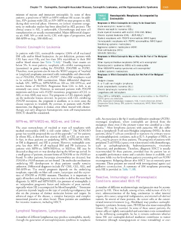
mixture of mature and immature eosinophils. In some of these TABLE Hematopoietic Neoplasms Accompanied by
patients, a prodrome of MDS or MPN without HE occurs. In addi- 71.8B Eosinophilia
tion, F/P+ patients with CEL or F/P+ MPN-eo may progress to AEL
during their terminal phase. However, no (other) recurrent cytoge- Neoplasms in Which Eosinophils Are Likely To Be Clonal Cells
netic or molecular marker has been identified in AEL patients. The Acute eosinophilic leukemia (AEL)
clinical course in AEL is unfavorable. Chemotherapy and stem cell Chronic eosinophilic leukemia (CEL)
transplantation are usually recommended. Major differential diagno- Acute myeloid leukemia with inv(16) (FAB AML M4eo)
+
ses are AML M4-eo with inv16, CEL with signs of progression, and Chronic myeloid leukemia (CML – BCR-ABL1 )
SM-AHN-eo (e.g., SM-AML-eo). Myeloid neoplasms with PDGFR abnormalities (WHO types)
Hematopoietic neoplasms with FGFR1 abnormalities (WHO types)
Smoldering systemic mastocytosis
Chronic Eosinophilic Leukemia Aggressive systemic mastocytosis (ASM)
Mast cell leukemia (MCL)
In patients with CEL, eosinophils comprise ≥30% of all nucleated SM-AHN (SM-CEL)
BM cells and/or blood leukocytes. As per definition, patients with Neoplasms in Which Eosinophils May or May Not Be Part of The Malignant
CEL have overt HE N and less than 20% myeloblasts in their BM Clone
3
and/or blood smears (see Table 71.8A). Usually, blast counts are Other myeloproliferative neoplasms (MPN) with eosinophilia a
below 5%. In most patients, neoplastic cells exhibit rearrangements Myelodysplastic syndromes (MDS) with eosinophilia
(mutations) in genes coding for PDGFRA, PDGFRB, or FGFR1. Other MDS/MPN overlap syndromes with eosinophilia a
Therefore, most patients also fit into the WHO category of “myeloid Indolent systemic mastocytosis
or lymphoid neoplasms associated with eosinophilia and abnormali- Neoplasms in Which Eosinophils Usually Are Not Part of the Malignant
ties of PDGFRA, PDGFRB, or FGFR1”. Other BM neoplasms need Clone
to be excluded by BM examination. Major differential diagnoses Hodgkin disease
include AEL, MPN-eo, MPN/MDS-eo, SM-eo, SM-AHN-eo, and B- or T-cell non-Hodgkin lymphoma
reactive forms of HE/HES. Progression from CEL to AEL is an Acute lymphoblastic leukemia (ALL)
extremely rare event. However, in untreated patients with PDGFR Chronic lymphocytic leukemia (CLL)
mutations and those with FGFR1 mutations, progression of CEL to Langerhans cell histiocytosis
AEL or even AML may occur. The prognosis in CEL depends largely a Other MPN or MPN/MDS: neoplasms where no abnormalities in the PDGFR or
on the molecular lesions detected. In patients with CEL exhibiting FGFR1 genes are detectable.
PDGFR mutations, the prognosis is excellent, as in most cases the AHN, Associated hematologic neoplasm; FAB, French–American–British
disease responds to imatinib. By contrast, in patients with FGFR1 Cooperative Study Group; SM, systemic mastocytosis; WHO, World Health
mutations, the diagnosis is dismal, even when treated with chemo- Organisation.
therapy and early allogeneic hematopoietic stem cell transplantation
(HSCT) should be considered.
cells. An exception is the 8p11 myeloproliferative syndrome (FGFR1-
MPN-eo, MPN/MDS-eo, MDS-eo, and SM-eo rearranged neoplasm), where eosinophils are derived from the
malignant clone even if the primary tumor was classified as a lym-
13
The exact nomenclature of myeloid or mast cell neoplasms with phoma. Most lymphoma patients with paracrine HE also suffer
20
marked eosinophilia (HE) is still under debate. The ICOG-EO from a (peripheral) T-cell non-Hodgkin lymphoma (NHL). In these
group has recently proposed the use of the appendix “-eo” for patients patients, clonal T cells are considered to represent the primary source
3
in whom HE N is detected but criteria of AEL or CEL are not met. of eosinophilopoietic cytokines, such as IL-5. A prephase of HES L or
In fact, in these patients an underlying MPN, MPN/MDS, MDS, HE US may be present in these patients. The prognosis of patients with
or SM is diagnosed, and HE is present. However, eosinophils com- peripheral T-cell NHLs is poor, even when treated with chemotherapy,
prise less than 30% of all nucleated BM and PB leukocytes. In such as cyclophosphamide, hydroxydaunomycin, vincristine
patients with MPN-eo, MPN/MDS-eo, or MDS-eo, HE may be (Oncovin), and prednisone. Therefore, allogeneic HSCT is usually
detected at diagnosis or may develop during the follow-up period. In recommended for these patients, provided that the patient has an
a small group of patients, mutant forms of PDGFRs or the FGFR are acceptable performance status and a suitable donor is available, and
found. In other patients, karyotype abnormalities are detected, but the same holds true for lymphoma patients presenting with an FGFR1
PDGFR or FGFR mutants are not found. The molecular mechanisms rearrangement. Relapsing disease after HSCT has an extremely poor
underlying HE development in these patients usually remains outcome. These patients are treated with investigational agents and
+
unknown. Differential diagnoses include Ph CML, CEL, AEL, and palliative cytoreduction. A summary of BM neoplasms presenting
SM-AHN-eo. The overall prognosis depends on the underlying with HE is provided in Table 71.8B.
neoplasm, especially on blast cell counts, karyotypes and the expres-
sion of PDGFR or FGFR1 mutants. Therefore, it is important to
apply all markers and diagnostic tests in order to define the exact final Reactive, Immunologic and Paraneoplastic
diagnosis. In MDS and MPN/MDS, the presence of eosinophilia is Conditions associated With HE
an independent prognostic factor and indicative of poor survival,
21
especially when HE is accompanied by blood basophilia. Treatment A number of different nonhematologic malignancies may be accom-
of patients depends largely on the type of underlying malignancy, but panied by HE. These include, among others, solid tumors of the GI
also on the presence of distinct molecular lesions and targets. In tract, adenocarcinomas of the lung, carcinomas in other internal
advanced stages of the disease, a complex karyotype and complex organs, skin cancer, urogenital malignancies, and stromal cell-derived
mutational patterns are often found. These patients are candidates tumors. In several of these patients, the cancer cells or the cancer-
for intensive treatment, including HSCT. related microenvironment (e.g., fibroblasts) may produce eosinophi-
lopoietic cytokines. In many cases, PB HE but also tissue eosinophilia
(tissue HE) may be recorded. In some of these cancer patients, the
Lymphoid Neoplasms, Lymphomas tumor cells are tightly surrounded and sometimes even outnumbered
by the infiltrating eosinophils. So far, it remains unknown whether
A number of different lymphomas may produce eosinophilia, mostly tissue HE and eosinophil-derived mediators contributes to tumor
through the generation of eosinophilopoietic cytokines in neoplastic formation (through neoangiogenesis or tissue remodeling) or even