
Page 1316 - Hematology_ Basic Principles and Practice ( PDFDrive )
P. 1316
1162 Part VII Hematologic Malignancies
microenvironment, including neoangiogenesis and BM fibrosis, is however, additional signs of myeloproliferation are found, consistent
usually detected in these patients. Depending on the underlying with a diagnosis of a MDS/MPN overlap syndrome or frank MPN.
19
neoplasm, other hematopoietic lineages may also be affected and may Another myeloid neoplasm that is often accompanied by HE is SM.
show typical morphological or phenotypic aberrations. Depending In fact, HE N is frequently seen in patients with advanced SM, includ-
on the underlying neoplasm, eosinophils may be mature or (rather) ing aggressive SM and mast cell leukemia (see Table 71.6). Most of
immature cells (Fig. 71.2B and C). The BM is also affected in patients these patients develop HE without HE-related organ damage (HES).
with reactive or idiopathic HES. However, in these patients eosino- In rare cases, lymphoid neoplasms may also present with HE. In these
phils are usually mature cells and no major alterations in the BM patients, a T-cell lymphoma is most commonly detected as underly-
microenvironment or abnormalities in other hematopoietic lineages ing disease during initial staging investigations or during follow-up.
are found. In most patients with T-cell lymphomas accompanied by HE, eosino-
Apart from eosinophilic leukemias, a number of different underly- phils are nonclonal cells. By contrast, in patients with fusions involv-
ing BM neoplasms may be identified, such as a MPN, myelodysplastic ing FGFR1, where stem cells are considered to give rise to malignant
syndrome (MDS), MDS/MPN overlap syndrome, acute myeloid cells, both eosinophils and lymphocytes are clonally involved. The
17
leukemia (AML), or, rarely, a lymphoid malignancy. In these prognosis in these patients is grave.
patients, various blood count abnormalities may be present, such as Several different concepts by which to classify eosinophil neo-
20
neutrophilia, monocytosis, basophilia, thrombocytosis, a left-shifted plasms have been proposed in the recent past. The WHO has
white blood count, or an increase in blasts. In some cases, anemia or/ classified eosinophil-related disorders according to the presence of
and thrombocytopenia with or without increased blast cells or dys- certain molecular lesions, including abnormalities in the PDGFR or
14
plasia is found, consistent with the diagnosis of an overt MDS, a FGFR genes (see Table 71.6). The advantage of this approach is that
MDS/MPN overlap syndrome, or AML. In rare cases, a stem cell it refers to molecular targets of therapy. As a result, therapeutic
neoplasm with involvement of both the myeloid and lymphoid decisions may be directly based on a WHO-based diagnosis. However,
lineage is detected. In these patients, an FGFR1-rearranged neoplasm independent of the clinical presentation and markers, the WHO
(previously referred to as 8p11 myeloproliferative syndrome or stem cell classification has to be complemented by a final histopathologic
13
leukemia/lymphoma syndrome) may be diagnosed. The prognosis is diagnosis, which in turn is based on thorough morphologic, histo-
poor in these patients. In some patients with CEL, a massive leuko- pathologic, and immunohistochemical studies of the BM. 3,17 Based
3
cytosis is found. In these cases, eosinophilia of more than 90% may on a proposal of the ICOG-EO group, the following underlying
be seen, and total leukocyte counts may exceed 50,000 or even neoplasms have to be delineated (see Table 71.8A):
3
100,000/mm . Extremely high leukocyte (eosinophil) counts are
more commonly seen in patients with HES N in the context of CEL
and are considered to be associated with a more unfavorable progno- Acute Eosinophilic Leukemia
sis. Blood smears from patients with HES R and HES I generally show
more or less normal, mature eosinophil morphologies, including This type of leukemia is defined by HE, more than 19% (≥20%)
typical bilobed nuclei and granule-rich cytoplasm. However, myeloblasts and greater than 29% (≥30%) eosinophils in BM smears
3
hypodense eosinophils and eosinophilic precursor cells may be (see Table 71.8A). Other classical types of MPN and CML must be
recorded, although less commonly, and eosinophils may also exhibit excluded. AEL is an extremely rare disease. Eosinophils in these
morphologic abnormalities, including nuclear hypersegmentation, patients may be quite immature, hypogranulated cells, or represent a
decreased size and/or numbers of secondary granules, and cytoplasmic
vacuolization. Mast cells may be increased in number in HES patients,
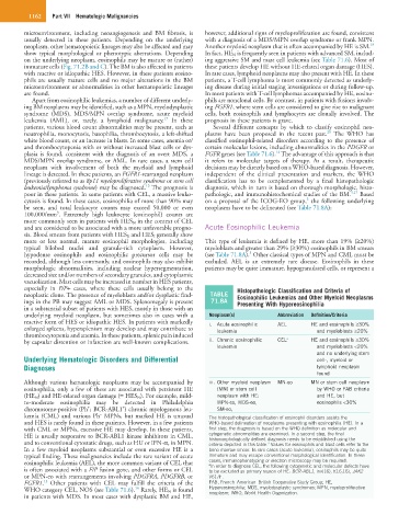
especially in F/P+ cases, where these cells usually belong to the Histopathologic Classification and Criteria of
neoplastic clone. The presence of myeloblasts and/or dysplastic find- TABLE Eosinophilic Leukemias and Other Myeloid Neoplasms
ings in the PB may suggest AML or MDS. Splenomegaly is present 71.8A Presenting With Hypereosinophilia
in a substantial subset of patients with HES, mostly in those with an
underlying myeloid neoplasm, but sometimes also in cases with a Neoplasm(s) Abbreviation Definition/Criteria
reactive form of HES or idiopathic HES. In patients with markedly i. Acute eosinophilic AEL HE and eosinophils ≥30%
enlarged spleens, hypersplenism may develop and may contribute to leukemia and myeloblasts ≥20%
thrombocytopenia and anemia. In these patients, splenic pain induced a
by capsular distention or infarction are well-known complications. ii. Chronic eosinophilic CEL HE and eosinophils ≥30%
leukemia and myeloblasts <20%
and no underlying stem
Underlying Hematologic Disorders and Differential cell-, myeloid or
Diagnoses lymphoid neoplasm
found
Although various hematologic neoplasms may be accompanied by iii. Other myeloid neoplasm MN-eo MN or stem cell neoplasm
eosinophilia, only a few of these are associated with persistent HE (MN) or stem cell by WHO or FAB criteria
(HE N ) and HE-related organ damage (= HES N ). For example, mild- neoplasm with HE: and HE, but
to-moderate eosinophilia may be detected in Philadelphia MPN-eo, MDS-eo, eosinophils <30%
+
+
chromosome-positive (Ph ; BCR-ABL1 ) chronic myelogenous leu- SM-eo,
−
kemia (CML) and various Ph MPNs, but marked HE is unusual The histopathological classification of eosinophil disorders assists the
and HES is rarely found in these patients. However, in a few patients WHO-based delineation of neoplasms presenting with eosinophilia (HE). In a
with CML or MPNs, excessive HE may develop. In these patients, first step, the diagnosis is based on the WHO definition as molecular and
HE is usually responsive to BCR-ABL1 kinase inhibitors in CML, cytogenetic abnormalities are examined. In a second step, the final
histomorphologically defined diagnosis needs to be established using the
and to conventional cytostatic drugs, such as HU or IFN-α, in MPN. criteria depicted in this table. Values for eosinophils and blast cells refer to the
3
In a few myeloid neoplasms substantial or even excessive HE is a bone marrow smear. In rare cases (acute leukemia), eosinophils may be quite
typical finding. These malignancies include the rare variant of acute immature and may escape conventional morphological identification. In these
eosinophilic leukemia (AEL), the more common variant of CEL that cases, immunophenotyping or electron microscopy may be required.
In order to diagnose CEL, the following cytogenetic and molecular defects have
a
is often associated with a F/P fusion gene, and other forms or CEL to be excluded as primary reason of HE: BCR-ABL1, inv(16), t(16;16), JAK2
or MPN-eo with rearrangements involving PDGFRA, PDGFRB, or V617F.
13
FGFR1. Other patients with CEL may fulfill the criteria of the FAB, French–American–British Cooperative Study Group; HE,
14
WHO category CEL, NOS (see Table 71.6). Rarely, HE N is found Hypereosinophilia; MDS, myelodysplastic syndromes; MPN, myeloproliferative
neoplasm; WHO, World Health Organization.
in patients with MDS. In most cases with dysplastic BM and HE,