
Page 1499 - Hematology_ Basic Principles and Practice ( PDFDrive )
P. 1499
Chapter 84 Malignant Lymphomas in Childhood 1331
Patients with a mass and less than 25% bone marrow (BM) lympho- Epidemiology
blasts are designated as having LBL, whereas patients with at least LBL constitutes 22% to 28% of childhood NHL. There is a 2 : 1 male
25% BM involvement have ALL. In contrast to ALL, more than 75% predominance for LBL, but the incidence of LBL remains constant
of patients with LBL demonstrate a precursor T-cell immunopheno- across the pediatric age group for both boys and girls. 14
type (T-LBL), with the remainder showing a precursor B-cell
immunophenotype (pB-LBL). 11–13 Pathobiology
LBL arises from precursor T or B lymphoblasts at varying stages of
differentiation. The morphology is similar to that of ALL, with
lymphoblasts of small or medium size and with scant cytoplasm,
round or convoluted nuclei, fine chromatin, and indistinct or small
TABLE Characteristics of Non-Hodgkin Lymphoma in Children nucleoli. Immunophenotyping shows terminal deoxynucleotidyl
84.1 transferase (TdT) positivity. T-LBL are usually positive for CD7 and
Proportion of surface or cytoplasmic CD3, with variable expression of CD2, CD5,
Cases in BFM CD1a, CD4, and CD8. Staining for TdT, CD3, myeloperoxidase,
Subtype Studies (%) 11 Phenotype Primary Site and a B-cell marker–like pax5 or CD79a are recommended parts of
15
a resource-saving diagnostic staining panel. CD10 expression is
Lymphoblastic 26 T cell; Mediastinum or more frequent in T-LBL (40%) than in T-ALL (less than 10%),
B cell head and neck; possibly related to maturational stage, with T-ALL more frequently
lymph nodes, skin, demonstrating an immature phenotype. 13,16,17 B-lineage markers are
soft tissue, bone
positive in pB-LBL. Unlike ALL, there are no known cytogenetic
Burkitt 49 B cell Abdomen or head prognostic factors for LBL. Recurrent cytogenetic anomalies are seen
and neck in about half of childhood T-ALL but have not been well-defined in
DLBCL 13 B cell Lymph nodes, T-LBL. Literature is scarce regarding typical chromosomal aberra-
mediastinum, tions for T-LBL. The commonest chromosomal translocations for
abdomen, head both T-LBL and T-ALL involve the T-cell receptor (TCR) gene loci
and neck at chromosome 14q11 or 7q34, resulting in the juxtaposition of an
ALCL 13 T cell Mediastinum, oncogenic partner gene with the regulatory region of one of the TCR
indeterminate abdomen, head gene loci and subsequent deregulation of a reciprocal partner gene,
and neck, bone, such as TAL1, MYC, HOXA gene cluster, and MYB. Certain molecu-
soft tissue, or lar genetic alterations were analyzed in relevant patient series of
skin pediatric T-LBL, including NOTCH1, FBXW7 mutations, altera-
tions of chromosome 9p containing CDKN2A/CDKN2B loci, and
ALCL, Anaplastic large-cell lymphoma; BFM, Berlin-Frankfurt-Münster; DLBCL, chromosome 6q. 18,19 Prospective systematic validation is required to
diffuse large B-cell lymphoma; Ig, immunoglobulin.
evaluate whether one of these markers or a combination of molecular
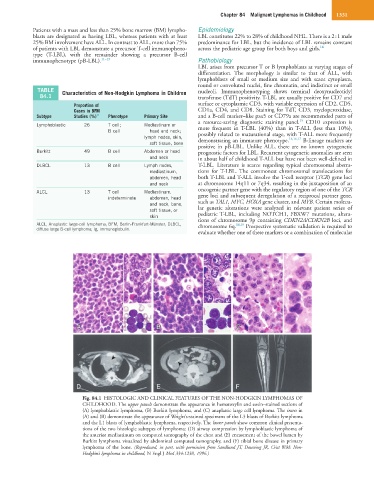
A A B B C C
D E F
Fig. 84.1 HISTOLOGIC AND CLINICAL FEATURES OF THE NON-HODGKIN LYMPHOMAS OF
CHILDHOOD. The upper panels demonstrate the appearance in hematoxylin and eosin–stained sections of
(A) lymphoblastic lymphoma, (B) Burkitt lymphoma, and (C) anaplastic large cell lymphoma. The insets in
(A) and (B) demonstrate the appearance of Wright’s-stained specimens of the L3 blasts of Burkitt lymphoma
and the L1 blasts of lymphoblastic lymphoma, respectively. The lower panels show common clinical presenta-
tions of the two histologic subtypes of lymphoma: (D) airway compression by lymphoblastic lymphoma of
the anterior mediastinum on computed tomography of the chest and (E) encasement of the bowel lumen by
Burkitt lymphoma visualized by abdominal computed tomography, and (F) tibial bone disease in primary
lymphoma of the bone. (Reproduced, in part, with permission from Sandlund JT, Downing JR, Crist WM: Non-
Hodgkin’s lymphoma in childhood, N Engl J Med 334:1238, 1996.)