
Page 1500 - Hematology_ Basic Principles and Practice ( PDFDrive )
P. 1500
1332 Part VII Hematologic Malignancies
26
markers are of stable prognostic relevance and can be used to optimize 80% in advanced-stage disease (Table 84.3). The Children’s Oncol-
current treatment stratification systems. ogy Group (COG) demonstrated that minimal disseminated disease
at diagnosis has prognostic value, as indicated by flow cytometric
Clinical Manifestations evidence of tumor cells in BM. In 99 children with T-LBL treated
The clinical features at presentation vary with primary site and extent in the A5971 study, 2-year event-free survival (EFS) was 68.1% ±
of disease spread. Almost all patients with T-LBL present with an 11.1% for patients with ≥1% T-LBL cells in BM by flow cytometry,
anterior mediastinal mass that may cause respiratory symptoms,
airway compromise, dysphagia, or superior vena cava syndrome (see
Fig. 84.1D). Pleural effusions are common (75%), and lymphade- TABLE
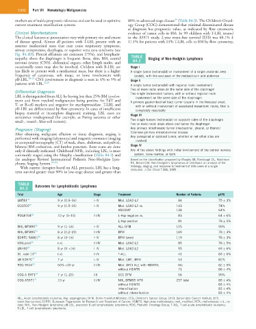
nopathy above the diaphragm is frequent. Bone, skin, BM, central 84.2 Staging of Non-Hodgkin Lymphoma
nervous system (CNS), abdominal organs, other lymph nodes, and
occasionally testes may also be involved. Children with B-LBL are Stage I
less likely to present with a mediastinal mass, but there is a higher A single tumor (extranodal) or involvement of a single anatomic area
frequency of cutaneous, soft tissue, or bone involvement with (nodal), with the exclusion of the mediastinum and abdomen
pB-LBL. 20–23 CNS involvement at diagnosis is seen in 4% to 5% of Stage II
patients with LBL. 22,24 A single tumor (extranodal) with regional node involvement
Two or more nodal areas on the same side of the diaphragm
Differential Diagnosis Two single (extranodal) tumors, with or without regional node
LBL is distinguished from ALL by having less than 25% BM involve- involvement on the same side of the diaphragm
ment and from myeloid malignancies being positive for TdT and A primary gastrointestinal tract tumor (usually in the ileocecal area),
T- or B-cell markers and negative for myeloperoxidase. T-LBL and with or without involvement of associated mesenteric nodes, that is
pB-LBL are differentiated by flow cytometry. In cases of insufficient completely resectable
biopsy material or incomplete diagnostic staining, LBL cases are Stage III
sometimes misdiagnosed (for example, as Ewing sarcoma or other
small-, round-, blue-cell tumors). Two single tumors (extranodal) on opposite sides of the diaphragm
Two or more nodal areas above and below the diaphragm
Prognosis (Staging) Any primary intrathoracic tumor (mediastinal, pleural, or thymic)
After obtaining malignant effusion or tissue diagnosis, staging is Extensive primary intraabdominal disease
performed with imaging (ultrasound and magnetic resonance imaging Any paraspinal or epidural tumor, whether or not other sites are
or computed tomography [CT] of neck, chest, abdomen, and pelvis), involved
bilateral BM evaluation, and lumbar puncture. Bone scans are done Stage IV
only if clinically indicated. Childhood NHL, including LBL, is most Any of the above findings with initial involvement of the central nervous
commonly staged using the Murphy classification (Table 84.2) and system, bone marrow, or both
the analogue Revised International Pediatric Non-Hodgkin Lym- Based on the classification proposed by Murphy SB, Fairclough DL, Hutchison
phoma Staging System. 25,25a RE, Berard CW: Non-Hodgkin’s lymphomas of childhood: an analysis of the
With current therapies based on ALL protocols, LBL has a long- histology, staging, and response to treatment of 338 cases at a single
term survival greater than 90% in low-stage disease and greater than institution. J Clin Oncol 7:186, 1989.
TABLE Outcomes for Lymphoblastic Lymphoma
84.3
Trial Age Stage Treatment Number of Patients pEFS
LMT81 31 9 yr (0.9–16) I–IV Mod. LSA2-L2 84 75 ± 3%
CCG502 32 9 yr (0.5–19) I–IV Mod. LSA2-L2 vs. 143 74%
ADCOMP 138 64%
POG8704 33 10 yr (5–15) III/IV L-Asp-negative vs. 83 64 ± 6%
L-Asp-positive 84 78 ± 5%
NHL-BFM90 26 9 yr (1–16) I–IV ALL-BFM 105 90%
NHL-BFM95 34 8 yr (0.2–19) III/IV BFM 169 78 ± 3%
EORTC 58881 35 8 yr (0–16) I–IV BFM-based 119 78 ± 3%
COG pilot 36 n.d. III/IV Mod. LSA2-L2 85 78 ± 5%
LNH92 37 8 yr (0–<16) I–IV Mod. LSA2-L2 55 69 ± 6%
St. Jude 13 38 n.d. III/V T-ALL 41 83 ± 6%
pB EORTC 22 7 yr I–IV Mod. LMT, BFM 53 82%
POG 9404 39 50% <10 yr III/IV Mod. DFCI ALL with HDMTX; 66; 82 ± 5%
without HDMTX 71 88 ± 4%
COG A 5971 40 7 yr (1–25) I/II CCG BFM 56 90%
COG A5971 41 10 yr III/IV NHL-BFM95 MTX 257 total 85 ± 4%
without HDMTX 83 ± 4%
intensification 83 ± 4%
without intensification 83 ± 4%
ALL, Acute lymphoblastic leukemia; Asp, asparaginase; BFM, Berlin-Frankfurt-Münster; CCG, Children’s Cancer Group; DFCI, Dana-Faber Cancer Institute; EFS,
event-free survival; EORTC, European Organisation for Research and Treatment of Cancer; HDMTX, high-dose methotrexate; mod, modified; MTX, methotrexate; n.d., no
data; NHL, Non-Hodgkin lymphoma; pB-LBL, precursor B-cell lymphoblastic lymphoma; POG, Pediatric Oncology Group; T-ALL, T-cell acute lymphoblastic leukemia;
T-LBL, T-cell lymphoblastic lymphoma.