
Page 1517 - Hematology_ Basic Principles and Practice ( PDFDrive )
P. 1517
1344 Part VII Hematologic Malignancies
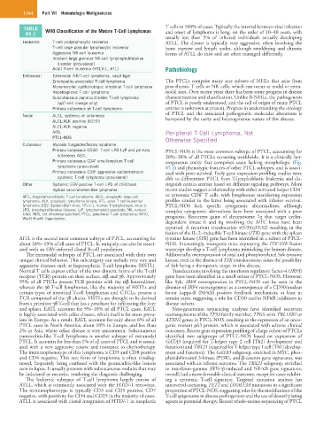
TABLE WHO Classification of the Mature T-Cell Lymphomas T cells in 100% of cases. Typically the interval between viral infection
85.1 and onset of lymphoma is long, on the order of 10–40 years, with
usually less than 5% of infected individuals actually developing
Leukemic T-cell prolymphocytic leukemia ATLL. The disease is typically very aggressive, often involving the
T-cell large granular lymphocytic leukemia bone marrow and lymph nodes, although smoldering and chronic
Aggressive NK-cell leukemia forms of ATLL do exist and are often managed differently.
Indolent large granular NK-cell lymphoproliferative
disorder (provisional)
Adult T-cell leukemia (HTLV-1, ATL) Pathobiology
Extranodal Extranodal NK/T-cell lymphoma, nasal type
Enteropathy-associated T-cell lymphoma The PTCLs comprise many rare subsets of NHLs that arise from
Monomorphic epitheliotropic intestinal T-cell lymphoma post-thymic T cells or NK cells, which can occur at nodal or extra-
Hepatosplenic T-cell lymphoma nodal sites. Over recent years there has been some progress in disease
Subcutaneous panniculitis-like T-cell lymphoma characterization and classification. Unlike B-NHLs, the pathogenesis
(αβT-cell lineage only) of PTCL is poorly understood, and the cell of origin of many PTCL
Primary cutaneous γδ T-cell lymphoma entities is unknown at present. Progress in understanding the etiology
of PTCL and the associated pathogenetic molecular alterations is
Nodal ALCL, systemic or cutaneous hampered by the rarity and heterogeneous nature of this disease.
ALCL:ALK positive [t(2;5)]
ALCL:ALK negative
AITL Peripheral T-Cell Lymphoma, Not
PTCL-NOS Otherwise Specified
Cutaneous Mycosis fungoides/Sézary syndrome
+
Primary cutaneous CD30 T-cell LPD LyP and primary PTCL-NOS is the most common subtype of PTCL, accounting for
cutaneous ALCL 20%–30% of all PTCLs occurring worldwide. It is a clinically het-
+
Primary cutaneous CD4 small/medium T-cell erogeneous entity that comprises cases lacking morphologic (Fig.
lymphoma (provisional) 85.3) and phenotypic features of other PTCL subtypes, and is associ-
Primary cutaneous CD8 aggressive epidermotropic ated with poor survival. Early gene expression profiling studies were
+
cytotoxic T-cell lymphoma (provisional) able to differentiate PTCL from T-lymphoblastic leukemia and dis-
Other Systemic EBV-positive T-cell LPD of childhood tinguish certain entities based on different signaling pathways. More
+
Hydroa vacciniforme–like lymphoma recent studies suggest a relationship with either activated helper CD4
+
AITL, Angioimmunoblastic T-cell lymphoma; ALCL, anaplastic large-cell or cytotoxic CD8 T cells, with lymphomas manifesting expression
lymphoma; ALK, anaplastic lymphoma kinase; ATL, adult T-cell leukemia/ profiles similar to the latter being associated with inferior survival.
lymphoma; EBV, Epstein-Barr virus; HTLV-1, human T-lymphotropic virus-1; PTCL-NOS lack specific cytogenetic abnormalities, although
LPD, lymphoproliferative disease; LyP, lymphomatoid papulosis; NK, natural complex cytogenetic aberrations have been associated with a poor
killer; NOS, not otherwise specified; PTCL, peripheral T-cell lymphoma; WHO, prognosis. Recurrent gains of chromosome 7q that target cyclin-
World Health Organization.
dependent kinase 6 and 8q involving the MYC locus have been
reported. A recurrent translocation t(5:9)(q33:32) resulting in the
fusion of the IL-2–inducible T-cell kinase (ITK) gene with the spleen
AITL is the second most common subtype of PTCL, accounting for tyrosine kinase (SYK) gene has been identified in a subset of PTCL-
about 18%–19% of all cases of PTCL. It, uniquely, can also be associ- NOS. Interestingly, transgenic mice expressing the ITK-SYK fusion
ated with an EBV-infected clonal B-cell population. transcript develop a T-cell lymphoma mimicking the human disease.
The extranodal subtypes of PTCL are associated with their own Additionally, overexpression of total and phosphorylated Syk tyrosine
unique clinical behavior. This subcategory can include very rare and kinase, even in the absence of SYK translocations, raises the possibility
aggressive diseases such as hepatosplenic T-cell lymphoma (HSTL). of Syk being a therapeutic target in this disease.
Normal T cells express either of the two dimeric forms of the T-cell Translocations involving the interferon regulatory factor-4 (IRF4)
receptor (TCR) protein on their surface, αβ and γδ. Approximately gene have been identified in a small subset of PTCL-NOS. However,
95% of all PTCLs possess TCR proteins with the αβ heterodimer, like Syk, IRF4 overexpression in PTCL-NOS can be seen in the
whereas the γδ T-cell lymphomas, like the majority of HSTLs and absence of IRF4 rearrangement as a consequence of a CD30/nuclear
certain types of intestinal T-cell lymphomas and CTCLs, possess a factor kappa-B (NFκB) positive feedback mechanism, at least in
TCR composed of the γδ chains. HSTLs are thought to be derived certain cases, suggesting a role for CD30 and/or NFκB inhibitors in
from a primitive γδ T-cell that has a penchant for infiltrating the liver disease subsets.
and spleen. EATL accounts for 5%–10% of all PTCL cases. EATL Next-generation sequencing analyses have identified recurrent
is highly associated with celiac disease, which itself is far more preva- rearrangements of the TP53 family member, TP63, with TBL1XR1 or
lent in Europe. As a result, EATL accounts for only about 6% of all ATXN1 genes in PTCL-NOS, resulting in the expression of an onco-
PTCL cases in North America, about 10% in Europe, and less than genic mutant p63 protein, which is associated with adverse clinical
2% in Asia, where celiac disease is very uncommon. Subcutaneous outcomes. Recent gene expression profiling of a large cohort of PTCLs
panniculitis-like T-cell lymphoma is another subtype of extranodal identified two subgroups of PTCL-NOS based on expression of
PTCL. It accounts for less than 1% of all cases of PTCL and is associ- GATA3 (required for T-helper type 2 cell [Th2] development and
ated with a very aggressive course and resistance to chemotherapy. function) and TBX21 (required for T-helper type 1 cell [Th1] develop-
The immunophenotype of this lymphoma is CD3 and CD8 positive ment and function). The GATA3 subgroup, enriched in MYC, phos-
and CD4 negative. This rare form of lymphoma is often misdiag- phatidylinositol 3-kinase (PI3K), and β-catenin gene signatures, was
nosed, frequently being confused with the panniculitic-like lesions associated with an inferior outcome. The TBX21 subgroup, enriched
seen in lupus. It usually presents with subcutaneous nodules that may in interferon-gamma (IFN-γ)-induced and NF-κB gene signatures,
be indurated or necrotic, rendering the diagnosis challenging. overall, had a more favorable clinical outcome, except for cases exhibit-
The leukemic subtypes of T-cell lymphoma largely consist of ing a cytotoxic T-cell signature. Targeted mutation analysis has
ATLL, which is commonly associated with the HTLV-1 retrovirus. uncovered coexisting TET2 and DNMT3A mutations in a significant
The immunophenotype is typically CD3 and CD5 positive, CD7 proportion of PTCL-NOS, suggesting roles for the modification of the
negative, with positivity for CD4 and CD25 in the majority of cases. T-cell epigenome in disease pathogenesis and the use of demethylating
ATLL is associated with clonal integration of HTLV-1 in neoplastic agents as potential therapy. Recent whole-exome sequencing of PTCL