
Page 1518 - Hematology_ Basic Principles and Practice ( PDFDrive )
P. 1518
Chapter 85 T-Cell Lymphomas 1345
Non-Hodgkin lymphoma
(NHL)
B-cell neoplasms T/NK-cell neoplasms
NHL neoplasm grouping Precursor lymphoid Mature T/NK-cell
neoplasms
neoplasms
Cutaneous Extranodal Nodal Leukemic
T-lymphoblastic Mycosis fungoides NKTCL Peripheral Adult T-cell
leukemia/lymphoma leukemia/
(MF) nasal type TCL-NOS
lymphoma
Anaplastic large Aggressive
2008 WHO classification of major subtypes syndrome Hepatosplenic Angioimmunoblastic prolymphocytic
Transformed
Enteropathy-
NK-cell
cell lymphoma
associated TCL
MF
leukemia
(ALK+/–)
T-cell
Sézary
TCL
TCL
leukemia
Subcutaneous
Primary cutaneous
+
lymphocytic
CD30 T-cell
leukemia
TCL
disorders panniculitis-like T-cell large granular
Primary cutaneous Aggressive
γ/δ TCL Indolent
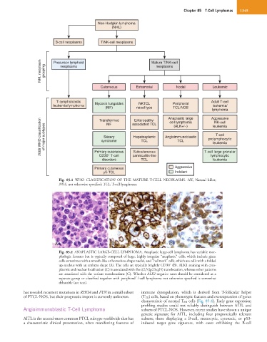
Fig. 85.1 WHO CLASSIFICATION OF THE MATURE T-CELL NEOPLASMS. NK, Natural killer;
NOS, not otherwise specified; TCL, T-cell lymphoma.
A A B B C C
Fig. 85.2 ANAPLASTIC LARGE-CELL LYMPHOMA. Anaplastic large-cell lymphoma has variable mor-
phologic features but is typically composed of large, highly irregular “anaplastic” cells, which include giant
cells sometimes with a wreath-like or horseshoe-shape nuclei, and “hallmark” cells, which are cells with a folded
+
up nucleus with an embryo shape (A). The cells are typically brightly CD30 (B). ALK1 staining with cyto-
plasmic and nuclear localization (C) is associated with the t(2;5)(p23;q35) translocation, whereas other patterns
are associated with the variant translocations (C). Whether ALK1-negative cases should be considered as a
separate group or classified together with peripheral T-cell lymphoma not otherwise specified is somewhat
debatable (see text).
has revealed recurrent mutations in RHOA and FYN in a small subset immune dysregulation, which is derived from T-follicular helper
of PTCL-NOS, but their prognostic import is currently unknown. (T FH) cells, based on phenotypic features and overexpression of genes
characteristic of normal T FH cells (Fig. 85.4). Early gene expression
profiling studies could not reliably distinguish between AITL and
Angioimmunoblastic T-Cell Lymphoma subsets of PTCL-NOS. However, recent studies have shown a unique
genetic signature for AITL, including four prognostically relevant
AITL is the second-most common PTCL subtype worldwide that has subsets; those displaying a B-cell, monocytic, cytotoxic, or p53-
a characteristic clinical presentation, often manifesting features of induced target gene signature, with cases exhibiting the B-cell